3DH7

Structure of T. thermophilus IDI-2 in complex with PPi


Experimental Data Snapshot

  • Method: X-RAY DIFFRACTION
  • Resolution: 2.97 Å
  • R-Value Free: 0.293 
  • R-Value Work: 0.211 
  • R-Value Observed: 0.215 

wwPDB Validation   3D Report Full Report


Ligand Structure Quality Assessment 


This is version 1.2 of the entry. See complete history


Literature

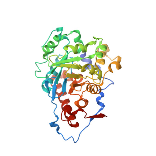
Crystal structure of type 2 isopentenyl diphosphate isomerase from Thermus thermophilus in complex with inorganic pyrophosphate

de Ruyck, J.Pouyez, J.Rothman, S.C.Poulter, D.Wouters, J.

(2008) Biochemistry 47: 9051-9053

  • DOI: https://doi.org/10.1021/bi801159x
  • Primary Citation of Related Structures:  
    3DH7

  • PubMed Abstract: 

    The N-terminal region is stabilized in the crystal structure of Thermus thermophilus type 2 isopentenyl diphosphate isomerase in complex with inorganic pyrophosphate, providing new insights about the active site and the catalytic mechanism of the enzyme. The PP i moiety is located near the conserved residues, H10, R97, H152, Q157, E158, and W219, and the flavin cofactor. The putative active site of isopentenyl diphosphate isomerase 2 provides interactions for stabilizing a carbocationic intermediate similar to those that stabilize the intermediate in the well-established protonation-deprotonation mechanism of isopentenyl diphosphate isomerase 1.


  • Organizational Affiliation

    Department of Chemistry, University of Namur, Namur, Belgium. jerome.deruyck@fundp.ac.be


Macromolecules
Find similar proteins by:  (by identity cutoff)  |  3D Structure
Entity ID: 1
MoleculeChains Sequence LengthOrganismDetailsImage
Isopentenyl-diphosphate delta-isomerase
A, B, C, D
332Thermus thermophilus HB8Mutation(s): 0 
Gene Names: TTHB110
UniProt
Find proteins for Q53W52 (Thermus thermophilus (strain ATCC 27634 / DSM 579 / HB8))
Explore Q53W52 
Go to UniProtKB:  Q53W52
Entity Groups  
Sequence Clusters30% Identity50% Identity70% Identity90% Identity95% Identity100% Identity
UniProt GroupQ53W52
Sequence Annotations
Expand
  • Reference Sequence
Experimental Data & Validation

Experimental Data

  • Method: X-RAY DIFFRACTION
  • Resolution: 2.97 Å
  • R-Value Free: 0.293 
  • R-Value Work: 0.211 
  • R-Value Observed: 0.215 
  • Space Group: P 32
Unit Cell:
Length ( Å )Angle ( ˚ )
a = 142.528α = 90
b = 142.528β = 90
c = 109.682γ = 120
Software Package:
Software NamePurpose
REFMACrefinement

Structure Validation

View Full Validation Report


Ligand Structure Quality Assessment 


Entry History 

Deposition Data

Revision History  (Full details and data files)

  • Version 1.0: 2008-09-23
    Type: Initial release
  • Version 1.1: 2011-07-13
    Changes: Version format compliance
  • Version 1.2: 2024-03-20
    Changes: Data collection, Database references, Derived calculations