


Crystal Structure of Drosophila Thioredoxin Reductase, C-terminal 8-residue truncation
Eckenroth, B.E., Hondal, R.J., Everse, S.J.To be published.
Experimental Data Snapshot
Entity ID: 1 | |||||
|---|---|---|---|---|---|
| Molecule | Chains | Sequence Length | Organism | Details | Image |
| Thioredoxin reductase 1, mitochondrial | 483 | Drosophila melanogaster | Mutation(s): 2 Gene Names: Trxr-1, GR, CG2151 EC: 1.8.1.9 | 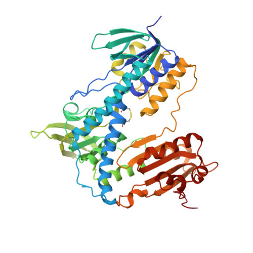 | |
UniProt | |||||
Find proteins for P91938 (Drosophila melanogaster) Explore P91938 Go to UniProtKB: P91938 | |||||
Entity Groups | |||||
| Sequence Clusters | 30% Identity50% Identity70% Identity90% Identity95% Identity100% Identity | ||||
| UniProt Group | P91938 | ||||
Sequence AnnotationsExpand | |||||
| |||||
| Ligands 2 Unique | |||||
|---|---|---|---|---|---|
| ID | Chains | Name / Formula / InChI Key | 2D Diagram | 3D Interactions | |
| FAD Query on FAD | C [auth A], F [auth B] | FLAVIN-ADENINE DINUCLEOTIDE C27 H33 N9 O15 P2 VWWQXMAJTJZDQX-UYBVJOGSSA-N |  | ||
| SO4 Query on SO4 | D [auth A], E [auth A], G [auth B], H [auth B] | SULFATE ION O4 S QAOWNCQODCNURD-UHFFFAOYSA-L |  | ||
| Length ( Å ) | Angle ( ˚ ) |
|---|---|
| a = 135.755 | α = 90 |
| b = 135.755 | β = 90 |
| c = 132.527 | γ = 90 |
| Software Name | Purpose |
|---|---|
| DENZO | data reduction |
| SCALEPACK | data scaling |
| MOLREP | phasing |
| REFMAC | refinement |
| PDB_EXTRACT | data extraction |
| Locally | data collection |
RCSB PDB (citation) is hosted by
RCSB PDB is a member of the

