3D3W

Structure of L-Xylulose Reductase with bound coenzyme, phosphate and hydroxide.


Experimental Data Snapshot

  • Method: X-RAY DIFFRACTION
  • Resolution: 1.87 Å
  • R-Value Free: 0.258 
  • R-Value Work: 0.204 
  • R-Value Observed: 0.207 

wwPDB Validation   3D Report Full Report


Ligand Structure Quality Assessment 


This is version 1.1 of the entry. See complete history


Literature

Structure/function analysis of a critical disulfide bond in the active site of L-xylulose reductase.

Zhao, H.T.Endo, S.Ishikura, S.Chung, R.Hogg, P.J.Hara, A.El-Kabbani, O.

(2009) Cell Mol Life Sci 66: 1570-1579

  • DOI: https://doi.org/10.1007/s00018-009-9065-y
  • Primary Citation of Related Structures:  
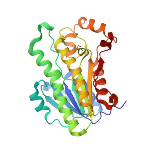
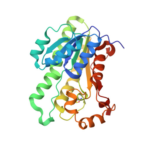
    3D3W

  • PubMed Abstract: 

    L-xylulose reductase (XR) is involved in water re-absorption and cellular osmoregulation. The crystal structure of human XR complemented with site-directed mutagenesis (Cys138Ala) indicated that the disulfide bond in the active site between Cys138 and Cys150 is unstable and may affect the reactivity of the enzyme. The effects of reducing agents on the activities of the wild-type and mutant enzymes indicated the reversibility of disulfide-bond formation, which resulted in three-fold decrease in catalytic efficiency. Furthermore, the addition of cysteine (>2 mM) inactivated human XR and was accompanied by a 10-fold decrease in catalytic efficiency. TOF-MS analysis of the inactivated enzyme showed the S-cysteinylation of Cys138 in the wild-type and Cys150 in the mutant enzymes. Thus, the action of human XR may be regulated by cellular redox conditions through reversible disulfide-bond formation and by S-cysteinylation.


  • Organizational Affiliation

    Medicinal Chemistry and Drug Action, Monash Institute of Pharmaceutical Sciences, 381 Royal Parade, Parkville, Victoria, 3052, Australia.


Macromolecules
Find similar proteins by:  (by identity cutoff)  |  3D Structure
Entity ID: 1
MoleculeChains Sequence LengthOrganismDetailsImage
L-xylulose reductase244Homo sapiensMutation(s): 0 
Gene Names: DCXR
EC: 1.1.1.10
UniProt & NIH Common Fund Data Resources
Find proteins for Q7Z4W1 (Homo sapiens)
Explore Q7Z4W1 
Go to UniProtKB:  Q7Z4W1
PHAROS:  Q7Z4W1
GTEx:  ENSG00000169738 
Entity Groups  
Sequence Clusters30% Identity50% Identity70% Identity90% Identity95% Identity100% Identity
UniProt GroupQ7Z4W1
Sequence Annotations
Expand
  • Reference Sequence
Find similar proteins by:  (by identity cutoff)  |  3D Structure
Entity ID: 2
MoleculeChains Sequence LengthOrganismDetailsImage
L-xylulose reductase244Homo sapiensMutation(s): 1 
Gene Names: DCXR
EC: 1.1.1.10
UniProt & NIH Common Fund Data Resources
Find proteins for Q7Z4W1 (Homo sapiens)
Explore Q7Z4W1 
Go to UniProtKB:  Q7Z4W1
PHAROS:  Q7Z4W1
GTEx:  ENSG00000169738 
Entity Groups  
Sequence Clusters30% Identity50% Identity70% Identity90% Identity95% Identity100% Identity
UniProt GroupQ7Z4W1
Sequence Annotations
Expand
  • Reference Sequence
Small Molecules
Modified Residues  1 Unique
IDChains TypeFormula2D DiagramParent
CSO
Query on CSO
A
L-PEPTIDE LINKINGC3 H7 N O3 SCYS
Experimental Data & Validation

Experimental Data

  • Method: X-RAY DIFFRACTION
  • Resolution: 1.87 Å
  • R-Value Free: 0.258 
  • R-Value Work: 0.204 
  • R-Value Observed: 0.207 
  • Space Group: P 21 21 2
Unit Cell:
Length ( Å )Angle ( ˚ )
a = 73.678α = 90
b = 87.476β = 90
c = 72.168γ = 90
Software Package:
Software NamePurpose
REFMACrefinement
HKL-2000data collection
HKL-2000data reduction
SCALEPACKdata scaling
MOLREPphasing

Structure Validation

View Full Validation Report


Ligand Structure Quality Assessment 


Entry History 

Deposition Data

Revision History  (Full details and data files)

  • Version 1.0: 2009-04-21
    Type: Initial release
  • Version 1.1: 2011-07-13
    Changes: Version format compliance