3CJJ

Crystal structure of human rage ligand-binding domain


Experimental Data Snapshot

  • Method: X-RAY DIFFRACTION
  • Resolution: 1.85 Å
  • R-Value Free: 0.240 
  • R-Value Work: 0.209 
  • R-Value Observed: 0.210 

wwPDB Validation   3D Report Full Report


This is version 1.2 of the entry. See complete history


Literature

Structural basis for ligand recognition and activation of RAGE.

Koch, M.Chitayat, S.Dattilo, B.M.Schiefner, A.Diez, J.Chazin, W.J.Fritz, G.

(2010) Structure 18: 1342-1352

  • DOI: https://doi.org/10.1016/j.str.2010.05.017
  • Primary Citation of Related Structures:  
    3CJJ

  • PubMed Abstract: 

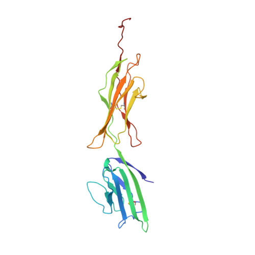
    The receptor for advanced glycation end products (RAGE) is a pattern recognition receptor involved in inflammatory processes and is associated with diabetic complications, tumor outgrowth, and neurodegenerative disorders. RAGE induces cellular signaling events upon binding of a variety of ligands, such as glycated proteins, amyloid-β, HMGB1, and S100 proteins. The X-ray crystal structure of the VC1 ligand-binding region of the human RAGE ectodomain was determined at 1.85 Å resolution. The VC1 ligand-binding surface was mapped onto the structure from titrations with S100B monitored by heteronuclear NMR spectroscopy. These NMR chemical shift perturbations were used as input for restrained docking calculations to generate a model for the VC1-S100B complex. Together, the arrangement of VC1 molecules in the crystal and complementary biochemical studies suggest a role for self-association in RAGE function. Our results enhance understanding of the functional outcomes of S100 protein binding to RAGE and provide insight into mechanistic models for how the receptor is activated.


  • Organizational Affiliation

    Department of Biology, University of Konstanz, 78457 Konstanz, Germany.


Macromolecules
Find similar proteins by:  (by identity cutoff)  |  3D Structure
Entity ID: 1
MoleculeChains Sequence LengthOrganismDetailsImage
Advanced glycosylation end product-specific receptor219Homo sapiensMutation(s): 0 
Gene Names: AGERRAGE
UniProt & NIH Common Fund Data Resources
Find proteins for Q15109 (Homo sapiens)
Explore Q15109 
Go to UniProtKB:  Q15109
PHAROS:  Q15109
GTEx:  ENSG00000204305 
Entity Groups  
Sequence Clusters30% Identity50% Identity70% Identity90% Identity95% Identity100% Identity
UniProt GroupQ15109
Sequence Annotations
Expand
  • Reference Sequence
Experimental Data & Validation

Experimental Data

  • Method: X-RAY DIFFRACTION
  • Resolution: 1.85 Å
  • R-Value Free: 0.240 
  • R-Value Work: 0.209 
  • R-Value Observed: 0.210 
  • Space Group: P 21 21 2
Unit Cell:
Length ( Å )Angle ( ˚ )
a = 74.85α = 90
b = 119.96β = 90
c = 28.89γ = 90
Software Package:
Software NamePurpose
XSCALEdata scaling
SHELXphasing
RESOLVEphasing
REFMACrefinement
PDB_EXTRACTdata extraction
SHELXDphasing

Structure Validation

View Full Validation Report


Entry History 

Deposition Data

Revision History  (Full details and data files)

  • Version 1.0: 2009-03-24
    Type: Initial release
  • Version 1.1: 2011-07-13
    Changes: Advisory, Version format compliance
  • Version 1.2: 2011-12-28
    Changes: Database references