3C84

Crystal structure of a complex of AChBP from aplysia californica and the neonicotinoid thiacloprid


Experimental Data Snapshot

  • Method: X-RAY DIFFRACTION
  • Resolution: 1.94 Å
  • R-Value Free: 0.215 
  • R-Value Work: 0.186 
  • R-Value Observed: 0.187 

wwPDB Validation   3D Report Full Report


Ligand Structure Quality Assessment 


This is version 1.2 of the entry. See complete history


Literature

Atomic interactions of neonicotinoid agonists with AChBP: molecular recognition of the distinctive electronegative pharmacophore.

Talley, T.T.Harel, M.Hibbs, R.E.Radic, Z.Tomizawa, M.Casida, J.E.Taylor, P.

(2008) Proc Natl Acad Sci U S A 105: 7606-7611

  • DOI: https://doi.org/10.1073/pnas.0802197105
  • Primary Citation of Related Structures:  
    3C79, 3C84

  • PubMed Abstract: 

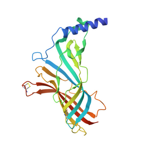
    Acetylcholine-binding proteins (AChBPs) from mollusks are suitable structural and functional surrogates of the nicotinic acetylcholine receptors when combined with transmembrane spans of the nicotinic receptor. These proteins assemble as a pentamer with identical ACh binding sites at the subunit interfaces and show ligand specificities resembling those of the nicotinic receptor for agonists and antagonists. A subset of ligands, termed the neonicotinoids, exhibit specificity for insect nicotinic receptors and selective toxicity as insecticides. AChBPs are of neither mammalian nor insect origin and exhibit a distinctive pattern of selectivity for the neonicotinoid ligands. We define here the binding orientation and determinants of differential molecular recognition for the neonicotinoids and classical nicotinoids by estimates of kinetic and equilibrium binding parameters and crystallographic analysis. Neonicotinoid complex formation is rapid and accompanied by quenching of the AChBP tryptophan fluorescence. Comparisons of the neonicotinoids imidacloprid and thiacloprid in the binding site from Aplysia californica AChBP at 2.48 and 1.94 A in resolution reveal a single conformation of the bound ligands with four of the five sites occupied in the pentameric crystal structure. The neonicotinoid electronegative pharmacophore is nestled in an inverted direction compared with the nicotinoid cationic functionality at the subunit interfacial binding pocket. Characteristic of several agonists, loop C largely envelops the ligand, positioning aromatic side chains to interact optimally with conjugated and hydrophobic regions of the neonicotinoid. This template defines the association of interacting amino acids and their energetic contributions to the distinctive interactions of neonicotinoids.


  • Organizational Affiliation

    Department of Pharmacology, Skaggs School of Pharmacy and Pharmaceutical Sciences, University of California at San Diego, La Jolla, CA 99093-0657, USA.


Macromolecules
Find similar proteins by:  (by identity cutoff)  |  3D Structure
Entity ID: 1
MoleculeChains Sequence LengthOrganismDetailsImage
Soluble acetylcholine receptor
A, B, C, D, E
227Aplysia californicaMutation(s): 0 
UniProt
Find proteins for Q8WSF8 (Aplysia californica)
Explore Q8WSF8 
Go to UniProtKB:  Q8WSF8
Entity Groups  
Sequence Clusters30% Identity50% Identity70% Identity90% Identity95% Identity100% Identity
UniProt GroupQ8WSF8
Sequence Annotations
Expand
  • Reference Sequence
Small Molecules
Ligands 3 Unique
IDChains Name / Formula / InChI Key2D Diagram3D Interactions
TH4
Query on TH4

Download Ideal Coordinates CCD File 
F [auth A],
H [auth B],
K [auth C],
O [auth D]
{(2Z)-3-[(6-chloropyridin-3-yl)methyl]-1,3-thiazolidin-2-ylidene}cyanamide
C10 H9 Cl N4 S
HOKKPVIRMVDYPB-UVTDQMKNSA-N
IPA
Query on IPA

Download Ideal Coordinates CCD File 
G [auth A]
I [auth B]
L [auth C]
M [auth C]
N [auth C]
G [auth A],
I [auth B],
L [auth C],
M [auth C],
N [auth C],
P [auth D],
Q [auth D]
ISOPROPYL ALCOHOL
C3 H8 O
KFZMGEQAYNKOFK-UHFFFAOYSA-N
MG
Query on MG

Download Ideal Coordinates CCD File 
J [auth C],
R [auth E]
MAGNESIUM ION
Mg
JLVVSXFLKOJNIY-UHFFFAOYSA-N
Binding Affinity Annotations 
IDSourceBinding Affinity
TH4 BindingDB:  3C84 Ki: 3.9 (nM) from 1 assay(s)
PDBBind:  3C84 Kd: 14 (nM) from 1 assay(s)
Binding MOAD:  3C84 Kd: 14 (nM) from 1 assay(s)
Experimental Data & Validation

Experimental Data

  • Method: X-RAY DIFFRACTION
  • Resolution: 1.94 Å
  • R-Value Free: 0.215 
  • R-Value Work: 0.186 
  • R-Value Observed: 0.187 
  • Space Group: P 21 21 21
Unit Cell:
Length ( Å )Angle ( ˚ )
a = 84.893α = 90
b = 116.726β = 90
c = 132.002γ = 90
Software Package:
Software NamePurpose
REFMACrefinement
HKL-2000data collection
DENZOdata reduction
SCALEPACKdata scaling
PHASERphasing

Structure Validation

View Full Validation Report


Ligand Structure Quality Assessment 


Entry History 

Deposition Data

Revision History  (Full details and data files)

  • Version 1.0: 2008-05-20
    Type: Initial release
  • Version 1.1: 2011-07-13
    Changes: Advisory, Version format compliance
  • Version 1.2: 2023-08-30
    Changes: Data collection, Database references, Derived calculations, Refinement description