3BPN

Crystal structure of the IL4-IL4R-IL13Ra ternary complex


Experimental Data Snapshot

  • Method: X-RAY DIFFRACTION
  • Resolution: 3.02 Å
  • R-Value Free: 0.304 
  • R-Value Work: 0.235 
  • R-Value Observed: 0.238 

wwPDB Validation   3D Report Full Report


Ligand Structure Quality Assessment 


This is version 1.3 of the entry. See complete history


Literature

Molecular and Structural Basis of Cytokine Receptor Pleiotropy in the Interleukin-4/13 System.

Laporte, S.L.Juo, Z.S.Vaclavikova, J.Colf, L.A.Qi, X.Heller, N.M.Keegan, A.D.Garcia, K.C.

(2008) Cell 132: 259-272

  • DOI: https://doi.org/10.1016/j.cell.2007.12.030
  • Primary Citation of Related Structures:  
    3BPL, 3BPN, 3BPO

  • PubMed Abstract: 

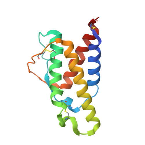
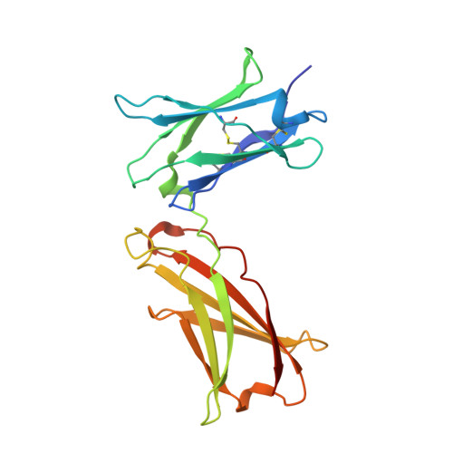
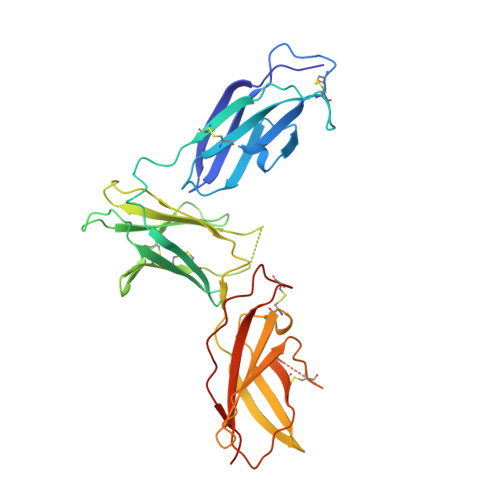
    Interleukin-4 and Interleukin-13 are cytokines critical to the development of T cell-mediated humoral immune responses, which are associated with allergy and asthma, and exert their actions through three different combinations of shared receptors. Here we present the crystal structures of the complete set of type I (IL-4R alpha/gamma(c)/IL-4) and type II (IL-4R alpha/IL-13R alpha1/IL-4, IL-4R alpha/IL-13R alpha1/IL-13) ternary signaling complexes. The type I complex reveals a structural basis for gamma(c)'s ability to recognize six different gamma(c)-cytokines. The two type II complexes utilize an unusual top-mounted Ig-like domain on IL-13R alpha1 for a novel mode of cytokine engagement that contributes to a reversal in the IL-4 versus IL-13 ternary complex assembly sequences, which are mediated through substantially different recognition chemistries. We also show that the type II receptor heterodimer signals with different potencies in response to IL-4 versus IL-13 and suggest that the extracellular cytokine-receptor interactions are modulating intracellular membrane-proximal signaling events.


  • Organizational Affiliation

    Howard Hughes Medical Institute, Department of Molecular and Cellular Physiology, Stanford University School of Medicine, Stanford, CA 94305, USA.


Macromolecules
Find similar proteins by:  (by identity cutoff)  |  3D Structure
Entity ID: 1
MoleculeChains Sequence LengthOrganismDetailsImage
Interleukin-4129Homo sapiensMutation(s): 0 
Gene Names: IL4
UniProt & NIH Common Fund Data Resources
Find proteins for P05112 (Homo sapiens)
Explore P05112 
Go to UniProtKB:  P05112
PHAROS:  P05112
GTEx:  ENSG00000113520 
Entity Groups  
Sequence Clusters30% Identity50% Identity70% Identity90% Identity95% Identity100% Identity
UniProt GroupP05112
Sequence Annotations
Expand
  • Reference Sequence
Find similar proteins by:  (by identity cutoff)  |  3D Structure
Entity ID: 2
MoleculeChains Sequence LengthOrganismDetailsImage
Interleukin-4 receptor alpha chain205Homo sapiensMutation(s): 4 
Gene Names: IL4R582J2.1IL4RA
UniProt & NIH Common Fund Data Resources
Find proteins for P24394 (Homo sapiens)
Explore P24394 
Go to UniProtKB:  P24394
PHAROS:  P24394
GTEx:  ENSG00000077238 
Entity Groups  
Sequence Clusters30% Identity50% Identity70% Identity90% Identity95% Identity100% Identity
UniProt GroupP24394
Sequence Annotations
Expand
  • Reference Sequence
Find similar proteins by:  (by identity cutoff)  |  3D Structure
Entity ID: 3
MoleculeChains Sequence LengthOrganismDetailsImage
Interleukin-13 receptor alpha-1 chain314Homo sapiensMutation(s): 0 
Gene Names: IL13RA1IL13RIL13RA
UniProt & NIH Common Fund Data Resources
Find proteins for P78552 (Homo sapiens)
Explore P78552 
Go to UniProtKB:  P78552
PHAROS:  P78552
GTEx:  ENSG00000131724 
Entity Groups  
Sequence Clusters30% Identity50% Identity70% Identity90% Identity95% Identity100% Identity
UniProt GroupP78552
Sequence Annotations
Expand
  • Reference Sequence
Small Molecules
Ligands 1 Unique
IDChains Name / Formula / InChI Key2D Diagram3D Interactions
NAG
Query on NAG

Download Ideal Coordinates CCD File 
D [auth B],
E [auth B]
2-acetamido-2-deoxy-beta-D-glucopyranose
C8 H15 N O6
OVRNDRQMDRJTHS-FMDGEEDCSA-N
Experimental Data & Validation

Experimental Data

  • Method: X-RAY DIFFRACTION
  • Resolution: 3.02 Å
  • R-Value Free: 0.304 
  • R-Value Work: 0.235 
  • R-Value Observed: 0.238 
  • Space Group: P 1 21 1
Unit Cell:
Length ( Å )Angle ( ˚ )
a = 61.64α = 90
b = 62.841β = 96.31
c = 115.125γ = 90
Software Package:
Software NamePurpose
REFMACrefinement
HKL-2000data collection
HKL-2000data reduction
SCALEPACKdata scaling
PHASERphasing

Structure Validation

View Full Validation Report


Ligand Structure Quality Assessment 


Entry History 

Deposition Data

  • Released Date: 2008-02-05 
  • Deposition Author(s): Garcia, K.C.

Revision History  (Full details and data files)

  • Version 1.0: 2008-02-05
    Type: Initial release
  • Version 1.1: 2011-07-13
    Changes: Advisory, Version format compliance
  • Version 1.2: 2020-07-29
    Type: Remediation
    Reason: Carbohydrate remediation
    Changes: Advisory, Data collection, Database references, Derived calculations, Structure summary
  • Version 1.3: 2021-10-20
    Changes: Advisory, Database references, Structure summary