Molecular Mechanism of ADP-Ribose Hydrolysis By Human NUDT5 From Structural and Kinetic Studies
Zha, M., Guo, Q., Zhang, Y., Yu, B., Ou, Y., Zhong, C., Ding, J.(2008) J Mol Biol 379: 568-578
- PubMed: 18462755
- DOI: https://doi.org/10.1016/j.jmb.2008.04.006
- Primary Citation of Related Structures:
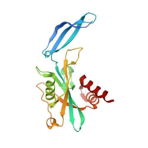
3BM4 - PubMed Abstract:
Human NUDT5 (hNUDT5) is an ADP-ribose (ADPR) pyrophosphatase (ADPRase) that plays important roles in controlling the intracellular levels of ADPR and preventing non-enzymatic ADP-ribosylation of proteins by hydrolyzing ADPR to AMP and ribose 5'-phosphate. We report the crystal structure of hNUDT5 in complex with a non-hydrolyzable ADPR analogue, alpha,beta-methyleneadenosine diphosphoribose, and three Mg(2+) ions representing the transition state of the enzyme during catalysis. Analysis of this structure and comparison with previously reported hNUDT5 structures identify key residues involved in substrate binding and catalysis. In the transition-state structure, three metal ions are bound at the active site and are coordinated by surrounding residues and water molecules. A conserved water molecule is at an ideal position for nucleophilic attack on the alpha-phosphate of ADPR. The side chain of Glu166 on loop L9 changes its conformation to interact with the conserved water molecule compared with that in the substrate-bound structure and appears to function as a catalytic base. Mutagenesis and kinetic studies show that Trp28 and Trp46 are important for the substrate binding; Arg51 is involved in both the substrate binding and the catalysis; and Glu112 and Glu116 of the Nudix motif, Glu166 on loop L9, and Arg111 are critical for the catalysis. The structural and biochemical data together reveal the molecular basis of the catalytic mechanism of ADPR hydrolysis by hNUDT5. Specifically, Glu166 functions as a catalytic base to deprotonate a conserved water molecule that acts as a nucleophile to attack the alpha-phosphate of ADPR, and three Mg(2+) ions are involved in the activation of the nucleophile and the binding of the substrate. Structural comparison of different ADPRases also suggests that most dimeric ADPRases may share a similar catalytic mechanism of ADPR hydrolysis.
Organizational Affiliation:
State Key Laboratory of Molecular Biology, Institute of Biochemistry and Cell Biology, Shanghai Institutes for Biological Sciences, Chinese Academy of Sciences, 320 Yue-Yang Road, Shanghai 200031, China.