3BJU

Crystal Structure of tetrameric form of human lysyl-tRNA synthetase


Experimental Data Snapshot

  • Method: X-RAY DIFFRACTION
  • Resolution: 2.31 Å
  • R-Value Free: 0.252 
  • R-Value Work: 0.194 
  • R-Value Observed: 0.197 

wwPDB Validation   3D Report Full Report


Ligand Structure Quality Assessment 


This is version 1.4 of the entry. See complete history


Literature

Crystal structure of tetrameric form of human lysyl-tRNA synthetase: Implications for multisynthetase complex formation

Guo, M.Ignatov, M.Musier-Forsyth, K.Schimmel, P.Yang, X.L.

(2008) Proc Natl Acad Sci U S A 105: 2331-2336

  • DOI: https://doi.org/10.1073/pnas.0712072105
  • Primary Citation of Related Structures:  
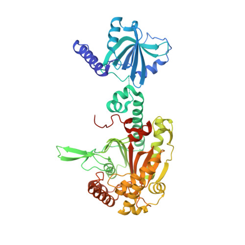
    3BJU

  • PubMed Abstract: 

    In mammals, many aminoacyl-tRNA synthetases are bound together in a multisynthetase complex (MSC) as a reservoir of procytokines and regulation molecules for functions beyond aminoacylation. The alpha(2) homodimeric lysyl-tRNA synthetase (LysRS) is tightly bound in the MSC and, under specific conditions, is secreted to trigger a proinflammatory response. Results by others suggest that alpha(2) LysRS is tightly bound into the core of the MSC with homodimeric beta(2) p38, a scaffolding protein that itself is multifunctional. Not understood is how the two dimeric proteins combine to make a presumptive alpha(2)beta(2) heterotetramer and, in particular, the location of the surfaces on LysRS that would accommodate the p38 interactions. Here we present a 2.3-A crystal structure of a tetrameric form of human LysRS. The relatively loose (as seen in solution) tetramer interface is assembled from two eukaryote-specific sequences, one in the catalytic- and another in the anticodon-binding domain. This same interface is predicted to provide unique determinants for interaction with p38. The analyses suggest how the core of the MSC is assembled and, more generally, that interactions and functions of synthetases can be built and regulated through dynamic protein-protein interfaces. These interfaces are created from small adaptations to what is otherwise a highly conserved (through evolution) polypeptide sequence.


  • Organizational Affiliation

    The Skaggs Institute for Chemical Biology and Department of Molecular Biology, The Scripps Research Institute, BCC-379, 10550 North Torrey Pines Road, La Jolla, CA 92037, USA.


Macromolecules
Find similar proteins by:  (by identity cutoff)  |  3D Structure
Entity ID: 1
MoleculeChains Sequence LengthOrganismDetailsImage
Lysyl-tRNA synthetase
A, B, C, D
521Homo sapiensMutation(s): 1 
Gene Names: KARSKIAA0070
EC: 6.1.1.6
UniProt & NIH Common Fund Data Resources
Find proteins for Q15046 (Homo sapiens)
Explore Q15046 
Go to UniProtKB:  Q15046
PHAROS:  Q15046
GTEx:  ENSG00000065427 
Entity Groups  
Sequence Clusters30% Identity50% Identity70% Identity90% Identity95% Identity100% Identity
UniProt GroupQ15046
Sequence Annotations
Expand
  • Reference Sequence
Small Molecules
Ligands 3 Unique
IDChains Name / Formula / InChI Key2D Diagram3D Interactions
ATP
Query on ATP

Download Ideal Coordinates CCD File 
I [auth A],
N [auth B],
S [auth C],
X [auth D]
ADENOSINE-5'-TRIPHOSPHATE
C10 H16 N5 O13 P3
ZKHQWZAMYRWXGA-KQYNXXCUSA-N
LYS
Query on LYS

Download Ideal Coordinates CCD File 
H [auth A],
M [auth B],
R [auth C],
W [auth D]
LYSINE
C6 H15 N2 O2
KDXKERNSBIXSRK-YFKPBYRVSA-O
CA
Query on CA

Download Ideal Coordinates CCD File 
E [auth A]
F [auth A]
G [auth A]
J [auth B]
K [auth B]
E [auth A],
F [auth A],
G [auth A],
J [auth B],
K [auth B],
L [auth B],
O [auth C],
P [auth C],
Q [auth C],
T [auth D],
U [auth D],
V [auth D]
CALCIUM ION
Ca
BHPQYMZQTOCNFJ-UHFFFAOYSA-N
Experimental Data & Validation

Experimental Data

  • Method: X-RAY DIFFRACTION
  • Resolution: 2.31 Å
  • R-Value Free: 0.252 
  • R-Value Work: 0.194 
  • R-Value Observed: 0.197 
  • Space Group: P 32
Unit Cell:
Length ( Å )Angle ( ˚ )
a = 152.013α = 90
b = 152.013β = 90
c = 108.838γ = 120
Software Package:
Software NamePurpose
DENZOdata reduction
SCALEPACKdata scaling
MOLREPphasing
DMphasing
REFMACrefinement
PDB_EXTRACTdata extraction

Structure Validation

View Full Validation Report


Ligand Structure Quality Assessment 


Entry History 

Deposition Data

Revision History  (Full details and data files)

  • Version 1.0: 2008-02-05
    Type: Initial release
  • Version 1.1: 2011-07-13
    Changes: Version format compliance
  • Version 1.2: 2017-10-25
    Changes: Refinement description
  • Version 1.3: 2021-10-20
    Changes: Database references, Derived calculations
  • Version 1.4: 2023-08-30
    Changes: Data collection, Refinement description