3B83

Computer-Based Redesign of a beta Sandwich Protein Suggests that Extensive Negative Design Is Not Required for De Novo beta Sheet Design.


Experimental Data Snapshot

  • Method: X-RAY DIFFRACTION
  • Resolution: 2.40 Å
  • R-Value Free: 0.290 
  • R-Value Work: 0.240 
  • R-Value Observed: 0.245 

wwPDB Validation   3D Report Full Report


This is version 1.3 of the entry. See complete history


Literature

Computer-Based Redesign of a beta Sandwich Protein Suggests that Extensive Negative Design Is Not Required for De Novo beta Sheet Design.

Hu, X.Wang, H.Ke, H.Kuhlman, B.

(2008) Structure 16: 1799-1805

  • DOI: https://doi.org/10.1016/j.str.2008.09.013
  • Primary Citation of Related Structures:  
    3B83

  • PubMed Abstract: 

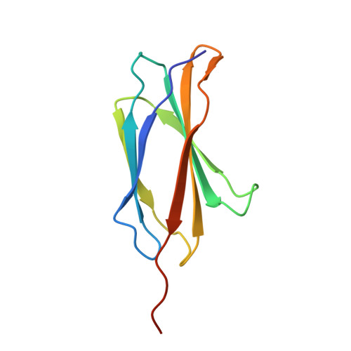
    The de novo design of globular beta sheet proteins remains largely an unsolved problem. It is unclear whether most designs are failing because the designed sequences do not have favorable energies in the target conformations or whether more emphasis should be placed on negative design, that is, explicitly identifying sequences that have poor energies when adopting undesired conformations. We tested whether we could redesign the sequence of a naturally occurring beta sheet protein, tenascin, with a design algorithm that does not include explicit negative design. Denaturation experiments indicate that the designs are significantly more stable than the wild-type protein and the crystal structure of one design closely matches the design model. These results suggest that extensive negative design is not required to create well-folded beta sandwich proteins. However, it is important to note that negative design elements may be encoded in the conformation of the protein backbone which was preserved from the wild-type protein.


  • Organizational Affiliation

    Department of Biochemistry and Biophysics, University of North Carolina at Chapel Hill, Chapel Hill, NC 27599-7260, USA.


Macromolecules
Find similar proteins by:  (by identity cutoff)  |  3D Structure
Entity ID: 1
MoleculeChains Sequence LengthOrganismDetailsImage
TEN-D3
A, B, C, D, E
A, B, C, D, E, F, G, H
100Homo sapiensMutation(s): 0 
Entity Groups  
Sequence Clusters30% Identity50% Identity70% Identity90% Identity95% Identity100% Identity
Sequence Annotations
Expand
  • Reference Sequence
Experimental Data & Validation

Experimental Data

  • Method: X-RAY DIFFRACTION
  • Resolution: 2.40 Å
  • R-Value Free: 0.290 
  • R-Value Work: 0.240 
  • R-Value Observed: 0.245 
  • Space Group: P 4 21 2
Unit Cell:
Length ( Å )Angle ( ˚ )
a = 126.329α = 90
b = 126.329β = 90
c = 134.661γ = 90
Software Package:
Software NamePurpose
ADSCdata collection
PHASERphasing
CNSrefinement
HKL-2000data reduction
HKL-2000data scaling

Structure Validation

View Full Validation Report


Entry History 

Deposition Data

Revision History  (Full details and data files)

  • Version 1.0: 2008-11-04
    Type: Initial release
  • Version 1.1: 2011-07-13
    Changes: Version format compliance
  • Version 1.2: 2013-06-05
    Changes: Structure summary
  • Version 1.3: 2023-08-30
    Changes: Data collection, Database references, Refinement description