


Crystal Structure of parkin ubiquitin-like domain R33Q mutant
Tomoo, K., Ikemiya, A., Amami, Y., In, Y., Ishida, T.To be published.
Experimental Data Snapshot
wwPDB Validation 3D Report Full Report

Entity ID: 1 | |||||
|---|---|---|---|---|---|
| Molecule | Chains | Sequence Length | Organism | Details | Image |
| E3 ubiquitin-protein ligase parkin | A [auth X] | 76 | Mus musculus | Mutation(s): 1 EC: 6.3.2 | 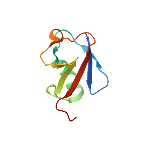 |
UniProt | |||||
Find proteins for Q9WVS6 (Mus musculus) Explore Q9WVS6 Go to UniProtKB: Q9WVS6 | |||||
Entity Groups | |||||
| Sequence Clusters | 30% Identity50% Identity70% Identity90% Identity95% Identity100% Identity | ||||
| UniProt Group | Q9WVS6 | ||||
Sequence AnnotationsExpand | |||||
| |||||
| Length ( Å ) | Angle ( ˚ ) |
|---|---|
| a = 45.79 | α = 90 |
| b = 45.79 | β = 90 |
| c = 65.98 | γ = 120 |
| Software Name | Purpose |
|---|---|
| REFMAC | refinement |
| PDB_EXTRACT | data extraction |
RCSB PDB (citation) is hosted by
RCSB PDB is a member of the

