Ligand specificity determined by differentially arranged common ligand-binding residues in bacterial amino acid chemoreceptors Tsr and Tar.
Tajima, H., Imada, K., Sakuma, M., Hattori, F., Nara, T., Kamo, N., Homma, M., Kawagishi, I.(2011) J Biol Chem 286: 42200-42210
- PubMed: 21979954
- DOI: https://doi.org/10.1074/jbc.M111.221887
- Primary Citation of Related Structures:
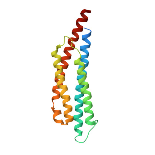
2D4U, 3ATP - PubMed Abstract:
Escherichia coli has closely related amino acid chemoreceptors with distinct ligand specificity, Tar for l-aspartate and Tsr for l-serine. Crystallography of the ligand-binding domain of Tar identified the residues interacting with aspartate, most of which are conserved in Tsr. However, swapping of the nonconserved residues between Tsr and Tar did not change ligand specificity. Analyses with chimeric receptors led us to hypothesize that distinct three-dimensional arrangements of the conserved ligand-binding residues are responsible for ligand specificity. To test this hypothesis, the structures of the apo- and serine-binding forms of the ligand-binding domain of Tsr were determined at 1.95 and 2.5 Å resolutions, respectively. Some of the Tsr residues are arranged differently from the corresponding aspartate-binding residues of Tar to form a high affinity serine-binding pocket. The ligand-binding pocket of Tsr was surrounded by negatively charged residues, which presumably exclude negatively charged aspartate molecules. We propose that all these Tsr- and Tar-specific features contribute to specific recognition of serine and aspartate with the arrangement of the side chain of residue 68 (Asn in Tsr and Ser in Tar) being the most critical.
Organizational Affiliation:
Division of Biological Science, Graduate School of Science, Nagoya University, Chikusa-ku, Nagoya 464-8602; Department of Frontier Bioscience, Hosei University, Koganei 184-8584; Research Center for Micro-Nano Technology, Hosei University, Koganei 184-8584.