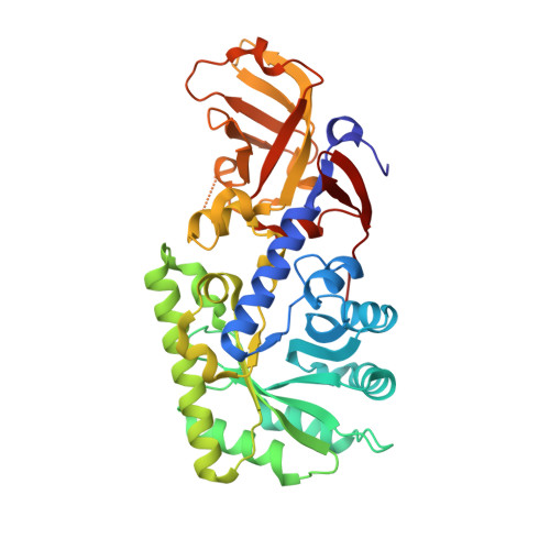
Crystal structure of a zinc-dependent D-serine dehydratase from chicken kidney
Tanaka, H., Senda, M., Venugopalan, N., Yamamoto, A., Senda, T., Ishida, T., Horiike, K.(2011) J Biol Chem 286: 27548-27558
- PubMed: 21676877
- DOI: https://doi.org/10.1074/jbc.M110.201160
- Primary Citation of Related Structures:
3ANU, 3ANV, 3AWN, 3AWO - PubMed Abstract:
D-serine is a physiological co-agonist of the N-methyl-D-aspartate receptor. It regulates excitatory neurotransmission, which is important for higher brain functions in vertebrates. In mammalian brains, D-amino acid oxidase degrades D-serine. However, we have found recently that in chicken brains the oxidase is not expressed and instead a D-serine dehydratase degrades D-serine. The primary structure of the enzyme shows significant similarities to those of metal-activated D-threonine aldolases, which are fold-type III pyridoxal 5'-phosphate (PLP)-dependent enzymes, suggesting that it is a novel class of D-serine dehydratase. In the present study, we characterized the chicken enzyme biochemically and also by x-ray crystallography. The enzyme activity on D-serine decreased 20-fold by EDTA treatment and recovered nearly completely by the addition of Zn(2+). None of the reaction products that would be expected from side reactions of the PLP-D-serine Schiff base were detected during the >6000 catalytic cycles of dehydration, indicating high reaction specificity. We have determined the first crystal structure of the D-serine dehydratase at 1.9 Å resolution. In the active site pocket, a zinc ion that coordinates His(347) and Cys(349) is located near the PLP-Lys(45) Schiff base. A theoretical model of the enzyme-D-serine complex suggested that the hydroxyl group of D-serine directly coordinates the zinc ion, and that the ε-NH(2) group of Lys(45) is a short distance from the substrate Cα atom. The α-proton abstraction from D-serine by Lys(45) and the elimination of the hydroxyl group seem to occur with the assistance of the zinc ion, resulting in the strict reaction specificity.
Organizational Affiliation:
Department of Biochemistry and Molecular Biology, Shiga University of Medical Science, Seta, Ohtsu, Shiga 520-2192, Japan.