3ACA

Crystal structure of human NUDT5 complexed with 8-oxo-dADP and manganese


Experimental Data Snapshot

  • Method: X-RAY DIFFRACTION
  • Resolution: 2.05 Å
  • R-Value Free: 0.247 
  • R-Value Work: 0.199 
  • R-Value Observed: 0.201 

wwPDB Validation   3D Report Full Report


Ligand Structure Quality Assessment 


This is version 1.4 of the entry. See complete history


Literature

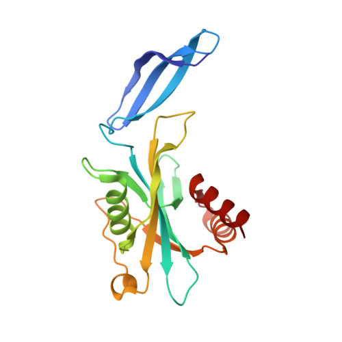
Diverse substrate recognition and hydrolysis mechanisms of human NUDT5

Arimori, T.Tamaoki, H.Nakamura, T.Kamiya, H.Ikemizu, S.Takagi, Y.Ishibashi, T.Harashima, H.Sekiguchi, M.Yamagata, Y.

(2011) Nucleic Acids Res 39: 8972-8983

  • DOI: https://doi.org/10.1093/nar/gkr575
  • Primary Citation of Related Structures:  
    3AC9, 3ACA, 3L85

  • PubMed Abstract: 

    Human NUDT5 (hNUDT5) hydrolyzes various modified nucleoside diphosphates including 8-oxo-dGDP, 8-oxo-dADP and ADP-ribose (ADPR). However, the structural basis of the broad substrate specificity remains unknown. Here, we report the crystal structures of hNUDT5 complexed with 8-oxo-dGDP and 8-oxo-dADP. These structures reveal an unusually different substrate-binding mode. In particular, the positions of two phosphates (α and β phosphates) of substrate in the 8-oxo-dGDP and 8-oxo-dADP complexes are completely inverted compared with those in the previously reported hNUDT5-ADPR complex structure. This result suggests that the nucleophilic substitution sites of the substrates involved in hydrolysis reactions differ despite the similarities in the chemical structures of the substrates and products. To clarify this hypothesis, we employed the isotope-labeling method and revealed that 8-oxo-dGDP is attacked by nucleophilic water at Pβ, whereas ADPR is attacked at Pα. This observation reveals that the broad substrate specificity of hNUDT5 is achieved by a diversity of not only substrate recognition, but also hydrolysis mechanisms and leads to a novel aspect that enzymes do not always catalyze the reaction of substrates with similar chemical structures by using the chemically equivalent reaction site.


  • Organizational Affiliation

    Graduate School of Pharmaceutical Sciences, Kumamoto University, Kumamoto 862-0973, Japan.


Macromolecules
Find similar proteins by:  (by identity cutoff)  |  3D Structure
Entity ID: 1
MoleculeChains Sequence LengthOrganismDetailsImage
ADP-sugar pyrophosphatase
A, B
196Homo sapiensMutation(s): 0 
Gene Names: NUDT5HSPC115
EC: 3.6.1.13
UniProt & NIH Common Fund Data Resources
Find proteins for Q9UKK9 (Homo sapiens)
Explore Q9UKK9 
Go to UniProtKB:  Q9UKK9
PHAROS:  Q9UKK9
GTEx:  ENSG00000165609 
Entity Groups  
Sequence Clusters30% Identity50% Identity70% Identity90% Identity95% Identity100% Identity
UniProt GroupQ9UKK9
Sequence Annotations
Expand
  • Reference Sequence
Experimental Data & Validation

Experimental Data

  • Method: X-RAY DIFFRACTION
  • Resolution: 2.05 Å
  • R-Value Free: 0.247 
  • R-Value Work: 0.199 
  • R-Value Observed: 0.201 
  • Space Group: C 1 2 1
Unit Cell:
Length ( Å )Angle ( ˚ )
a = 113.636α = 90
b = 40.701β = 121.64
c = 99.791γ = 90
Software Package:
Software NamePurpose
PHENIXrefinement
PDB_EXTRACTdata extraction
HKL-2000data collection
HKL-2000data reduction
HKL-2000data scaling
CNSphasing

Structure Validation

View Full Validation Report


Ligand Structure Quality Assessment 


Entry History 

Deposition Data

Revision History  (Full details and data files)

  • Version 1.0: 2011-01-12
    Type: Initial release
  • Version 1.1: 2011-07-13
    Changes: Version format compliance
  • Version 1.2: 2011-11-09
    Changes: Database references, Structure summary
  • Version 1.3: 2013-06-05
    Changes: Database references
  • Version 1.4: 2023-11-01
    Changes: Data collection, Database references, Derived calculations, Refinement description