X-ray structures of Bacillus pallidusd-arabinose isomerase and its complex with l-fucitol.
Takeda, K., Yoshida, H., Izumori, K., Kamitori, S.(2010) Biochim Biophys Acta 1804: 1359-1368
- PubMed: 20123133
- DOI: https://doi.org/10.1016/j.bbapap.2010.01.018
- Primary Citation of Related Structures:
3A9R, 3A9S, 3A9T - PubMed Abstract:
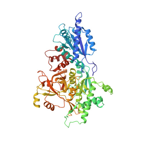
d-Arabinose isomerase (d-AI), also known as l-fucose isomerase (l-FI), catalyzes the aldose-ketose isomerization of d-arabinose to d-ribulose, and l-fucose to l-fuculose. Bacillus pallidus (B. pallidus) d-AI can catalyze isomerization of d-altrose to d-psicose, as well as d-arabinose and l-fucose. Three X-ray structures of B. pallidus d-AI in complexes with 2-methyl-2,4-pentadiol, glycerol and an inhibitor, l-fucitol, were determined at resolutions of 1.77, 1.60 and 2.60 A, respectively. B. pallidus d-AI forms a homo-hexamer, and one subunit has three domains of almost equal size; two Rossmann fold domains and a mimic of the (beta/alpha) barrel fold domain. A catalytic metal ion (Mn(2+)) was found in the active site coordinated by Glu342, Asp366 and His532, and an additional metal ion was found at the channel for the passage of a substrate coordinated by Asp453. The X-ray structures basically supported the ene-diol mechanism for the aldose-ketose isomerization by B. pallidus d-AI, as well as Escherichia coli (E. coli) l-FI, in which Glu342 and Asp366 facing each other at the catalytic metal ion transfer a proton from C2 to C1 and O1 to O2, acting as acid/base catalysts, respectively. However, considering the ionized state of Asp366, the catalytic reaction also possibly occurs through the negatively charged ene-diolate intermediate stabilized by the catalytic metal ion. A structural comparison with E. colil-FI showed that B. pallidus d-AI possibly interconverts between "open" and "closed" forms, and that the additional metal ion found in B. pallidus d-AI may help to stabilize the channel region.
Organizational Affiliation:
Life Science Research Center and Faculty of Medicine, Kagawa University, Miki-cho, Kita-gun, Kagawa 761-0793, Japan.