3A5E

Crystal structure of 5K RNase Sa


Experimental Data Snapshot

  • Method: X-RAY DIFFRACTION
  • Resolution: 1.60 Å
  • R-Value Free: 0.281 
  • R-Value Work: 0.229 
  • R-Value Observed: 0.231 

wwPDB Validation   3D Report Full Report


This is version 1.2 of the entry. See complete history


Literature

Urea denatured state ensembles contain extensive secondary structure that is increased in hydrophobic proteins

Nick Pace, C.Huyghues-Despointes, B.M.Fu, H.Takano, K.Scholtz, J.M.Grimsley, G.R.

(2010) Protein Sci 19: 929-943

  • DOI: https://doi.org/10.1002/pro.370
  • Primary Citation of Related Structures:  
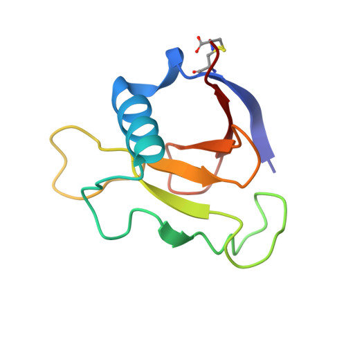
    3A5E

  • PubMed Abstract: 

    The goal of this article is to gain a better understanding of the denatured state ensemble (DSE) of proteins through an experimental and computational study of their denaturation by urea. Proteins unfold to different extents in urea and the most hydrophobic proteins have the most compact DSE and contain almost as much secondary structure as folded proteins. Proteins that unfold to the greatest extent near pH 7 still contain substantial amounts of secondary structure. At low pH, the DSE expands due to charge-charge interactions and when the net charge per residue is high, most of the secondary structure is disrupted. The proteins in the DSE appear to contain substantial amounts of polyproline II conformation at high urea concentrations. In all cases considered, including staph nuclease, the extent of unfolding by urea can be accounted for using the data and approach developed in the laboratory of Wayne Bolen (Auton et al., Proc Natl Acad Sci 2007; 104:15317-15323).


  • Organizational Affiliation

    Department of Biochemistry and Biophysics, Texas A&M University, College Station, Texas 77843, USA. nickpace@tamu.edu


Macromolecules
Find similar proteins by:  (by identity cutoff)  |  3D Structure
Entity ID: 1
MoleculeChains Sequence LengthOrganismDetailsImage
Guanyl-specific ribonuclease Sa96Kitasatospora aureofaciensMutation(s): 5 
Gene Names: rnaSA
EC: 3.1.27.3
UniProt
Find proteins for P05798 (Kitasatospora aureofaciens)
Explore P05798 
Go to UniProtKB:  P05798
Entity Groups  
Sequence Clusters30% Identity50% Identity70% Identity90% Identity95% Identity100% Identity
UniProt GroupP05798
Sequence Annotations
Expand
  • Reference Sequence
Experimental Data & Validation

Experimental Data

  • Method: X-RAY DIFFRACTION
  • Resolution: 1.60 Å
  • R-Value Free: 0.281 
  • R-Value Work: 0.229 
  • R-Value Observed: 0.231 
  • Space Group: P 21 21 21
Unit Cell:
Length ( Å )Angle ( ˚ )
a = 51.339α = 90
b = 46.241β = 90
c = 36.744γ = 90
Software Package:
Software NamePurpose
HKL-2000data collection
AMoREphasing
REFMACrefinement
HKL-2000data reduction
HKL-2000data scaling

Structure Validation

View Full Validation Report


Entry History 

Deposition Data

  • Released Date: 2010-08-04 
  • Deposition Author(s): Takano, K.

Revision History  (Full details and data files)

  • Version 1.0: 2010-08-04
    Type: Initial release
  • Version 1.1: 2011-07-13
    Changes: Version format compliance
  • Version 1.2: 2023-11-01
    Changes: Data collection, Database references, Refinement description