3A29

Crystal structure of human liver FBPase in complex with tricyclic inhibitor


Experimental Data Snapshot

  • Method: X-RAY DIFFRACTION
  • Resolution: 2.60 Å
  • R-Value Free: 0.247 
  • R-Value Work: 0.204 
  • R-Value Observed: 0.208 

wwPDB Validation   3D Report Full Report


Ligand Structure Quality Assessment 


This is version 1.3 of the entry. See complete history


Literature

Synthesis, SAR, and X-ray structure of tricyclic compounds as potent FBPase inhibitors

Tsukada, T.Takahashi, M.Takemoto, T.Kanno, O.Yamane, T.Kawamura, S.Nishi, T.

(2009) Bioorg Med Chem Lett 19: 5909-5912

  • DOI: https://doi.org/10.1016/j.bmcl.2009.08.081
  • Primary Citation of Related Structures:  
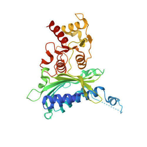
    3A29

  • PubMed Abstract: 

    With the aim of discovering a novel class of fructose-1,6-bisphosphatase (FBPase) inhibitors, a series of compounds based on tricyclic scaffolds was synthesized. Extensive SAR studies led to the finding of 8l with an IC50 value of 0.013 microM against human FBPase. An X-ray crystallographic study revealed that 8l bound at AMP binding sites of human liver FBPase with hydrogen bonding interactions similar to AMP.


  • Organizational Affiliation

    Medicinal Chemistry Research Laboratories I, Daiichi Sankyo Co., Ltd, 1-2-58 Hiromachi, Shinagawa-ku, Tokyo 140-8710, Japan.


Macromolecules
Find similar proteins by:  (by identity cutoff)  |  3D Structure
Entity ID: 1
MoleculeChains Sequence LengthOrganismDetailsImage
Fructose-1,6-bisphosphatase 1
A, B, C, D
337Homo sapiensMutation(s): 0 
Gene Names: FBP1FBP
EC: 3.1.3.11
UniProt & NIH Common Fund Data Resources
Find proteins for P09467 (Homo sapiens)
Explore P09467 
Go to UniProtKB:  P09467
PHAROS:  P09467
GTEx:  ENSG00000165140 
Entity Groups  
Sequence Clusters30% Identity50% Identity70% Identity90% Identity95% Identity100% Identity
UniProt GroupP09467
Sequence Annotations
Expand
  • Reference Sequence
Small Molecules
Binding Affinity Annotations 
IDSourceBinding Affinity
2T0 PDBBind:  3A29 IC50: 13 (nM) from 1 assay(s)
BindingDB:  3A29 IC50: 13 (nM) from 1 assay(s)
Experimental Data & Validation

Experimental Data

  • Method: X-RAY DIFFRACTION
  • Resolution: 2.60 Å
  • R-Value Free: 0.247 
  • R-Value Work: 0.204 
  • R-Value Observed: 0.208 
  • Space Group: P 21 21 21
Unit Cell:
Length ( Å )Angle ( ˚ )
a = 67.289α = 90
b = 82.851β = 90
c = 277.253γ = 90
Software Package:
Software NamePurpose
CNXrefinement
DENZOdata reduction
SCALEPACKdata scaling
X-PLORphasing

Structure Validation

View Full Validation Report


Ligand Structure Quality Assessment 


Entry History 

Deposition Data

Revision History  (Full details and data files)

  • Version 1.0: 2009-10-06
    Type: Initial release
  • Version 1.1: 2011-07-13
    Changes: Version format compliance
  • Version 1.2: 2017-10-11
    Changes: Refinement description
  • Version 1.3: 2023-11-01
    Changes: Data collection, Database references, Derived calculations, Refinement description