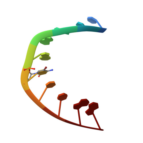
Nine polymorphic crystal structures of d(CCGGGCCCGG), d(CCGGGCCm5CGG), d(Cm5CGGGCCm5CGG) and d(CCGGGCC(Br)5CGG) in three different conformations: effects of spermine binding and methylation on the bending and condensation of A-DNA.
Tippin, D.B., Sundaralingam, M.(1997) J Mol Biol 267: 1171-1185
- PubMed: 9150405
- DOI: https://doi.org/10.1006/jmbi.1997.0945
- Primary Citation of Related Structures:
318D, 319D, 320D, 321D, 322D, 323D, 324D, 325D, 326D - PubMed Abstract:
The A-DNA decamer d(CCGGGCCm5CGG) crystallizes in the presence of spermine in three polymorphic forms and with one duplex in the asymmetric unit: hexagonal (P6(1)), unit cell of 55.0 A x 55.0 A x 45.9 A; orthorhombic (P2(1)2(1)2(1)), unit cell of 24.8 A x 44.6 A x 48.0 A, and a second orthorhombic (P2(1)2(1)2(1)), unit cell of 23.6 A x 40.8 A x 43.4 A. The reduction in cell volume among the three different forms is accompanied by a large reduction in solvent content (67% versus 40% versus 24%) and a significant reduction in volume per base-pair (2005 A(3) versus 1325 A(3) versus 1048 A(3)). There is also a concomitant increase in the number of bound spermine molecules per duplex (0 versus 1 versus 2) as well as an increase in DNA bending (10 degrees versus 16 degrees versus 31 degrees), which correspond to major groove widths of 8.0 A versus 4.5 A versus 1.3 A, respectively. The P6(1) crystal form, which represents a new space group for A-DNA decamers, supports one of the most hydrated and extended DNA duplexes to date, while the second orthorhombic form supports one of the least-hydrated and most-condensed non-Z-DNA duplexes. The unmethylated analogue d(CCGGGCCCGG), the double-methyl derivative d(Cm5CGGGCCm5CGG) and the bromine derivative d(CCGGGCC(Br)5CGG) also crystallize in at least two of the aforementioned conformations, and all nine crystal structures were determined. We report, in detail, on the three crystal structures of d(CCGGGCCm5CGG) and the effects of methylation and spermine binding on A-DNA conformation.
Organizational Affiliation:
Department of Chemistry, The Ohio State University, Columbus 43210-1002, USA.