2ZFJ

Crystal Structure of the Kif1A Motor Domain during Mg release: Mg-releasing Transition-1


Experimental Data Snapshot

  • Method: X-RAY DIFFRACTION
  • Resolution: 3.20 Å
  • R-Value Free: 0.298 
  • R-Value Work: 0.267 
  • R-Value Observed: 0.267 

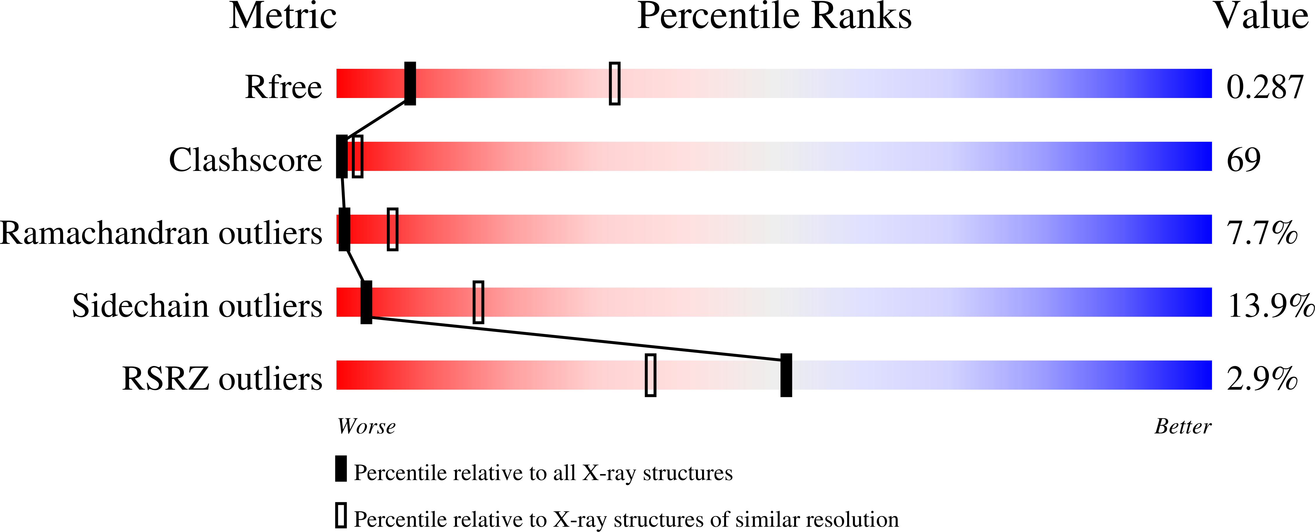
wwPDB Validation   3D Report Full Report


Ligand Structure Quality Assessment 


This is version 1.3 of the entry. See complete history


Literature

Structural model for strain-dependent microtubule activation of Mg-ADP release from kinesin.

Nitta, R.Okada, Y.Hirokawa, N.

(2008) Nat Struct Mol Biol 15: 1067-1075

  • DOI: https://doi.org/10.1038/nsmb.1487
  • Primary Citation of Related Structures:  
    2ZFI, 2ZFJ, 2ZFK, 2ZFL, 2ZFM

  • PubMed Abstract: 

    Mg-ADP release is considered to be a crucial process for the regulation and motility of kinesin. To gain insight into the structural basis of this process, we solved the atomic structures of kinesin superfamily protein-1A (KIF1A) during and after Mg(2+) release. On the basis of new structural and mutagenesis data, we propose a model mechanism for microtubule activation of Mg-ADP release from KIF1A. In our model, a specific interaction between loop L7 of KIF1A and beta-tubulin reconfigures the KIF1A active site by shifting the relative positions of switches I and II. This leads to the sequential release of a group of water molecules that sits over the Mg(2+) in the active site, followed by Mg(2+) and finally the ADP. We further propose that this set of events is linked to a strain-dependent docking of the neck linker to the motor core, which produces a two-step power stroke.


  • Organizational Affiliation

    Department of Cell Biology and Anatomy, University of Tokyo, Graduate School of Medicine, 7-3-1 Hongo, Bunkyo-ku, Tokyo, 113-0033, Japan.


Macromolecules
Find similar proteins by:  (by identity cutoff)  |  3D Structure
Entity ID: 1
MoleculeChains Sequence LengthOrganismDetailsImage
Kinesin-like protein KIF1A, Kinesin heavy chain isoform 5C366Mus musculusMutation(s): 0 
UniProt
Find proteins for P33173 (Mus musculus)
Explore P33173 
Go to UniProtKB:  P33173
Entity Groups  
Sequence Clusters30% Identity50% Identity70% Identity90% Identity95% Identity100% Identity
UniProt GroupP33173
Sequence Annotations
Expand
  • Reference Sequence
Small Molecules
Ligands 2 Unique
IDChains Name / Formula / InChI Key2D Diagram3D Interactions
ADP
Query on ADP

Download Ideal Coordinates CCD File 
C [auth A]ADENOSINE-5'-DIPHOSPHATE
C10 H15 N5 O10 P2
XTWYTFMLZFPYCI-KQYNXXCUSA-N
MG
Query on MG

Download Ideal Coordinates CCD File 
B [auth A]MAGNESIUM ION
Mg
JLVVSXFLKOJNIY-UHFFFAOYSA-N
Experimental Data & Validation

Experimental Data

  • Method: X-RAY DIFFRACTION
  • Resolution: 3.20 Å
  • R-Value Free: 0.298 
  • R-Value Work: 0.267 
  • R-Value Observed: 0.267 
  • Space Group: P 21 21 21
Unit Cell:
Length ( Å )Angle ( ˚ )
a = 40.142α = 90
b = 51.202β = 90
c = 157.012γ = 90
Software Package:
Software NamePurpose
CNSrefinement
ADSCdata collection
HKL-2000data reduction
HKL-2000data scaling
CNSphasing

Structure Validation

View Full Validation Report


Ligand Structure Quality Assessment 


Entry History 

Deposition Data

Revision History  (Full details and data files)

  • Version 1.0: 2008-09-23
    Type: Initial release
  • Version 1.1: 2011-07-13
    Changes: Version format compliance
  • Version 1.2: 2017-08-23
    Changes: Source and taxonomy
  • Version 1.3: 2023-11-01
    Changes: Data collection, Database references, Refinement description