2Z8W

Structure of an IgNAR-AMA1 complex


Experimental Data Snapshot

  • Method: X-RAY DIFFRACTION
  • Resolution: 2.45 Å
  • R-Value Free: 0.282 
  • R-Value Work: 0.189 
  • R-Value Observed: 0.198 

wwPDB Validation   3D Report Full Report


This is version 1.3 of the entry. See complete history


Literature

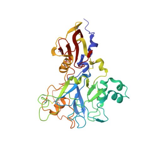
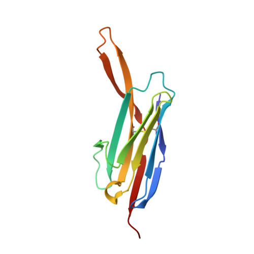
Structure of an IgNAR-AMA1 Complex: Targeting a Conserved Hydrophobic Cleft Broadens Malarial Strain Recognition

Henderson, K.A.Streltsov, V.A.Coley, A.M.Dolezal, O.Hudson, P.J.Batchelor, A.H.Gupta, A.Bai, T.Murphy, V.J.Anders, R.F.Foley, M.Nuttall, S.D.

(2007) Structure 15: 1452-1466

  • DOI: https://doi.org/10.1016/j.str.2007.09.011
  • Primary Citation of Related Structures:  
    2Z8V, 2Z8W

  • PubMed Abstract: 

    Apical membrane antigen 1 (AMA1) is essential for invasion of erythrocytes and hepatocytes by Plasmodium parasites and is a leading malarial vaccine candidate. Although conventional antibodies to AMA1 can prevent such invasion, extensive polymorphisms within surface-exposed loops may limit the ability of these AMA1-induced antibodies to protect against all parasite genotypes. Using an AMA1-specific IgNAR single-variable-domain antibody, we performed targeted mutagenesis and selection against AMA1 from three P. falciparum strains. We present cocrystal structures of two antibody-AMA1 complexes which reveal extended IgNAR CDR3 loops penetrating deep into a hydrophobic cleft on the antigen surface and contacting residues conserved across parasite species. Comparison of a series of affinity-enhancing mutations allowed dissection of their relative contributions to binding kinetics and correlation with inhibition of erythrocyte invasion. These findings provide insights into mechanisms of single-domain antibody binding, and may enable design of reagents targeting otherwise cryptic epitopes in pathogen antigens.


  • Organizational Affiliation

    CSIRO Molecular and Health Technologies, 343 Royal Parade, Parkville 3052, Australia.


Macromolecules
Find similar proteins by:  (by identity cutoff)  |  3D Structure
Entity ID: 1
MoleculeChains Sequence LengthOrganismDetailsImage
Apical membrane antigen 1
A, B
335Plasmodium falciparum 3D7Mutation(s): 0 
UniProt
Find proteins for Q7KQK5 (Plasmodium falciparum (isolate 3D7))
Explore Q7KQK5 
Go to UniProtKB:  Q7KQK5
Entity Groups  
Sequence Clusters30% Identity50% Identity70% Identity90% Identity95% Identity100% Identity
UniProt GroupQ7KQK5
Sequence Annotations
Expand
  • Reference Sequence
Find similar proteins by:  (by identity cutoff)  |  3D Structure
Entity ID: 2
MoleculeChains Sequence LengthOrganismDetailsImage
New antigen receptor variable domain
C, D
116Orectolobus maculatusMutation(s): 2 
UniProt
Find proteins for Q6X1E6 (Orectolobus maculatus)
Explore Q6X1E6 
Go to UniProtKB:  Q6X1E6
Entity Groups  
Sequence Clusters30% Identity50% Identity70% Identity90% Identity95% Identity100% Identity
UniProt GroupQ6X1E6
Sequence Annotations
Expand
  • Reference Sequence
Experimental Data & Validation

Experimental Data

  • Method: X-RAY DIFFRACTION
  • Resolution: 2.45 Å
  • R-Value Free: 0.282 
  • R-Value Work: 0.189 
  • R-Value Observed: 0.198 
  • Space Group: P 31
Unit Cell:
Length ( Å )Angle ( ˚ )
a = 76.48α = 90
b = 76.48β = 90
c = 140.978γ = 120
Software Package:
Software NamePurpose
REFMACrefinement
ADSCdata collection
HKL-2000data reduction
HKL-2000data scaling
PHASERphasing

Structure Validation

View Full Validation Report


Entry History 

Deposition Data

Revision History  (Full details and data files)

  • Version 1.0: 2007-11-27
    Type: Initial release
  • Version 1.1: 2011-07-13
    Changes: Advisory, Source and taxonomy, Version format compliance
  • Version 1.2: 2021-11-10
    Changes: Database references
  • Version 1.3: 2023-11-01
    Changes: Data collection, Refinement description