2Z33

Solution structure of the DNA complex of PhoB DNA-binding/transactivation Domain


Experimental Data Snapshot

  • Method: SOLUTION NMR
  • Conformers Calculated: 100 
  • Conformers Submitted: 10 
  • Selection Criteria: structures with the least restraint violations, structures with the lowest energy 

wwPDB Validation   3D Report Full Report


This is version 1.2 of the entry. See complete history


Literature

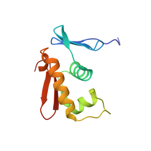
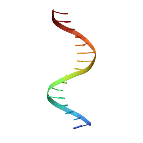
Water-mediated interactions between DNA and PhoB DNA-binding/transactivation domain: NMR-restrained molecular dynamics in explicit water environment.

Yamane, T.Okamura, H.Ikeguchi, M.Nishimura, Y.Kidera, A.

(2008) Proteins 71: 1970-1983

  • DOI: https://doi.org/10.1002/prot.21874
  • Primary Citation of Related Structures:  
    2Z33

  • PubMed Abstract: 

    The solution structure of the complex between the transcription factor PhoB DNA-binding/transactivation domain and DNA was determined by NMR spectroscopy and simulated annealing in a periodic boundary box of explicit water with the particle mesh Ewald method. The refined structures provided better convergence and better local geometry compared with the structures determined in vacuum. The hydrogen bond interactions between the PhoB domain and DNA in the aqueous environment were fully formed. The complex structure was found to be very similar to the crystal structure, particularly at the PhoB-DNA interface, much more so than expected from the vacuum structure. These results indicate the importance of the proper treatment of electrostatic and hydration influences in describing protein-DNA interactions. The hydration structures observed for the refined structures contained most of the crystal waters as a subset. We observed that various water-mediated PhoB-DNA interactions contributed to the molecular recognition between PhoB and DNA.


  • Organizational Affiliation

    International Graduate School of Arts and Sciences, Yokohama City University, Yokohama, Japan.


Macromolecules

Find similar proteins by:  (by identity cutoff)  |  3D Structure
Entity ID: 3
MoleculeChains Sequence LengthOrganismDetailsImage
Phosphate regulon transcriptional regulatory protein phoBC [auth A]104Escherichia coliMutation(s): 0 
Gene Names: phoB
UniProt
Find proteins for P0AFJ5 (Escherichia coli (strain K12))
Explore P0AFJ5 
Go to UniProtKB:  P0AFJ5
Entity Groups  
Sequence Clusters30% Identity50% Identity70% Identity90% Identity95% Identity100% Identity
UniProt GroupP0AFJ5
Sequence Annotations
Expand
  • Reference Sequence

Find similar nucleic acids by:  Sequence   |   3D Structure  

Entity ID: 1
MoleculeChains LengthOrganismImage
5'-D(*AP*CP*TP*GP*TP*CP*AP*TP*AP*AP*AP*TP*CP*TP*GP*T)-3'A [auth B]16N/A
Sequence Annotations
Expand
  • Reference Sequence

Find similar nucleic acids by:  Sequence   |   3D Structure  

Entity ID: 2
MoleculeChains LengthOrganismImage
5'-D(*AP*CP*AP*GP*AP*TP*TP*TP*AP*TP*GP*AP*CP*AP*GP*T)-3'B [auth C]16N/A
Sequence Annotations
Expand
  • Reference Sequence
Experimental Data & Validation

Experimental Data

  • Method: SOLUTION NMR
  • Conformers Calculated: 100 
  • Conformers Submitted: 10 
  • Selection Criteria: structures with the least restraint violations, structures with the lowest energy 

Structure Validation

View Full Validation Report


Entry History 

Deposition Data

Revision History  (Full details and data files)

  • Version 1.0: 2008-04-22
    Type: Initial release
  • Version 1.1: 2011-07-13
    Changes: Version format compliance
  • Version 1.2: 2022-03-16
    Changes: Database references, Derived calculations