


Crystal structure of pyridoxine biosynthesis protein from Methanocaldococcus jannaschii
Manzoku, M., Ebihara, A., Chen, L., Fu, Z.-Q., Chrzas, J., Wang, B.-C., Yokoyama, S., Kuramitsu, S.To be published.
Experimental Data Snapshot
wwPDB Validation 3D Report Full Report

Entity ID: 1 | |||||
|---|---|---|---|---|---|
| Molecule | Chains | Sequence Length | Organism | Details | Image |
| Pyridoxal biosynthesis lyase pdxS | 330 | Methanocaldococcus jannaschii DSM 2661 | Mutation(s): 0 EC: 4 | 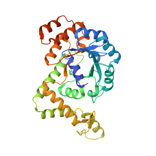 | |
UniProt | |||||
Find proteins for Q58090 (Methanocaldococcus jannaschii (strain ATCC 43067 / DSM 2661 / JAL-1 / JCM 10045 / NBRC 100440)) Explore Q58090 Go to UniProtKB: Q58090 | |||||
Entity Groups | |||||
| Sequence Clusters | 30% Identity50% Identity70% Identity90% Identity95% Identity100% Identity | ||||
| UniProt Group | Q58090 | ||||
Sequence AnnotationsExpand | |||||
| |||||
| Ligands 1 Unique | |||||
|---|---|---|---|---|---|
| ID | Chains | Name / Formula / InChI Key | 2D Diagram | 3D Interactions | |
| CL Query on CL | D [auth A] E [auth A] F [auth A] G [auth A] H [auth B] | CHLORIDE ION Cl VEXZGXHMUGYJMC-UHFFFAOYSA-M |  | ||
| Length ( Å ) | Angle ( ˚ ) |
|---|---|
| a = 106.054 | α = 90 |
| b = 170.797 | β = 90 |
| c = 173.334 | γ = 90 |
| Software Name | Purpose |
|---|---|
| CNS | refinement |
| HKL-2000 | data reduction |
| HKL-2000 | data scaling |
| MOLREP | phasing |
RCSB PDB (citation) is hosted by
RCSB PDB is a member of the

