


Trapping of the uric acid substrate in the crystal structure of urate oxidase from Arthrobacter globiformis
Juan, E.C.M., Hossain, M.T., Hoque, M.M., Yamamoto, T., Imamura, S., Suzuki, K., Sekiguchi, T., Takenaka, A.To be published.
Experimental Data Snapshot
Entity ID: 1 | |||||
|---|---|---|---|---|---|
| Molecule | Chains | Sequence Length | Organism | Details | Image |
| Uricase | 302 | Arthrobacter globiformis | Mutation(s): 0 EC: 1.7.3.3 | 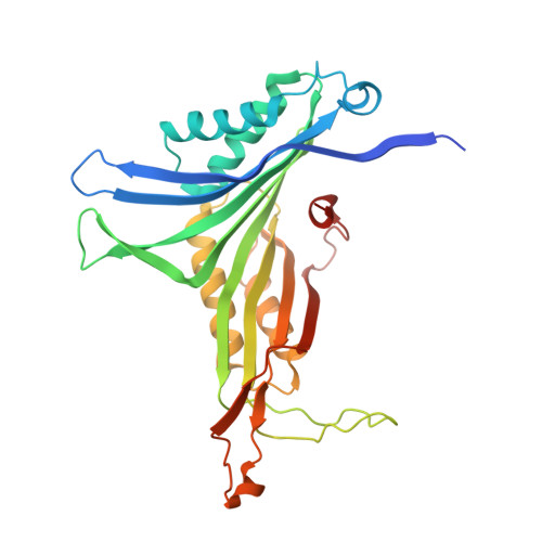 | |
UniProt | |||||
Find proteins for D0VWQ1 (Arthrobacter globiformis) Explore D0VWQ1 Go to UniProtKB: D0VWQ1 | |||||
Entity Groups | |||||
| Sequence Clusters | 30% Identity50% Identity70% Identity90% Identity95% Identity100% Identity | ||||
| UniProt Group | D0VWQ1 | ||||
Sequence AnnotationsExpand | |||||
| |||||
| Ligands 1 Unique | |||||
|---|---|---|---|---|---|
| ID | Chains | Name / Formula / InChI Key | 2D Diagram | 3D Interactions | |
| 1AL Query on 1AL | I [auth A] J [auth B] K [auth B] L [auth D] M [auth E] | ALLANTOATE ION C4 H7 N4 O4 NUCLJNSWZCHRKL-UHFFFAOYSA-M |  | ||
| Length ( Å ) | Angle ( ˚ ) |
|---|---|
| a = 85.498 | α = 90 |
| b = 122.942 | β = 90 |
| c = 284.784 | γ = 90 |
| Software Name | Purpose |
|---|---|
| REFMAC | refinement |
| HKL-2000 | data collection |
| HKL-2000 | data reduction |
| HKL-2000 | data scaling |
| AMoRE | phasing |
RCSB PDB (citation) is hosted by
RCSB PDB is a member of the

