2YAN

Crystal structure of the second glutaredoxin domain of human TXNL2


Experimental Data Snapshot

  • Method: X-RAY DIFFRACTION
  • Resolution: 1.90 Å
  • R-Value Free: 0.212 
  • R-Value Work: 0.185 
  • R-Value Observed: 0.186 

wwPDB Validation   3D Report Full Report


Ligand Structure Quality Assessment 


This is version 1.4 of the entry. See complete history


Literature

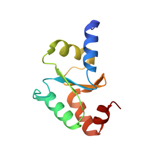
Crystal Structure of the Second Glutaredoxin Domain of Human Txnl2

Vollmar, M.Johansson, C.Cocking, R.Muniz, J.R.C.Krojer, T.Allerston, C.von Delft, F.Bountra, C.Arrowsmith, C.H.Weigelt, J.Edwards, A.Oppermann, U.

To be published.

Macromolecules
Find similar proteins by:  (by identity cutoff)  |  3D Structure
Entity ID: 1
MoleculeChains Sequence LengthOrganismDetailsImage
GLUTAREDOXIN-3
A, B
105Homo sapiensMutation(s): 0 
UniProt & NIH Common Fund Data Resources
Find proteins for O76003 (Homo sapiens)
Explore O76003 
Go to UniProtKB:  O76003
PHAROS:  O76003
GTEx:  ENSG00000108010 
Entity Groups  
Sequence Clusters30% Identity50% Identity70% Identity90% Identity95% Identity100% Identity
UniProt GroupO76003
Sequence Annotations
Expand
  • Reference Sequence
Small Molecules
Ligands 5 Unique
IDChains Name / Formula / InChI Key2D Diagram3D Interactions
GSH
Query on GSH

Download Ideal Coordinates CCD File 
J [auth A],
N [auth B]
GLUTATHIONE
C10 H17 N3 O6 S
RWSXRVCMGQZWBV-WDSKDSINSA-N
SO4
Query on SO4

Download Ideal Coordinates CCD File 
H [auth A]SULFATE ION
O4 S
QAOWNCQODCNURD-UHFFFAOYSA-L
EDO
Query on EDO

Download Ideal Coordinates CCD File 
C [auth A]
D [auth A]
E [auth A]
F [auth A]
K [auth B]
C [auth A],
D [auth A],
E [auth A],
F [auth A],
K [auth B],
L [auth B],
M [auth B]
1,2-ETHANEDIOL
C2 H6 O2
LYCAIKOWRPUZTN-UHFFFAOYSA-N
FE
Query on FE

Download Ideal Coordinates CCD File 
G [auth A]FE (III) ION
Fe
VTLYFUHAOXGGBS-UHFFFAOYSA-N
CL
Query on CL

Download Ideal Coordinates CCD File 
I [auth A]CHLORIDE ION
Cl
VEXZGXHMUGYJMC-UHFFFAOYSA-M
Experimental Data & Validation

Experimental Data

  • Method: X-RAY DIFFRACTION
  • Resolution: 1.90 Å
  • R-Value Free: 0.212 
  • R-Value Work: 0.185 
  • R-Value Observed: 0.186 
  • Space Group: P 32 2 1
Unit Cell:
Length ( Å )Angle ( ˚ )
a = 118.317α = 90
b = 118.317β = 90
c = 58.84γ = 120
Software Package:
Software NamePurpose
REFMACrefinement
XDSdata reduction
SCALAdata scaling
PHASERphasing

Structure Validation

View Full Validation Report


Ligand Structure Quality Assessment 


Entry History 

Revision History  (Full details and data files)

  • Version 1.0: 2011-11-30
    Type: Initial release
  • Version 1.1: 2012-01-18
    Changes: Other
  • Version 1.2: 2012-12-05
    Changes: Database references
  • Version 1.3: 2018-01-24
    Changes: Database references
  • Version 1.4: 2023-12-20
    Changes: Data collection, Database references, Derived calculations, Other, Refinement description