2Y0E

BceC and the final step of UGDs reaction


Experimental Data Snapshot

  • Method: X-RAY DIFFRACTION
  • Resolution: 1.75 Å
  • R-Value Free: 0.197 
  • R-Value Work: 0.163 
  • R-Value Observed: 0.165 

wwPDB Validation   3D Report Full Report


Ligand Structure Quality Assessment 


This is version 1.2 of the entry. See complete history


Literature

Structure of Burkholderia Cepacia Udp-Glucose Dehydrogenase (Ugd) Bcec and Role of Tyr10 in Final Hydrolysis of Ugd Thioester Intermediate.

Rocha, J.Popescu, A.O.Borges, P.Mil-Homens, D.Moreira, L.M.Sa-Correia, I.Fialho, A.M.Frazao, C.

(2011) J Bacteriol 193: 3978

  • DOI: https://doi.org/10.1128/JB.01076-10
  • Primary Citation of Related Structures:  
    2Y0C, 2Y0D, 2Y0E

  • PubMed Abstract: 

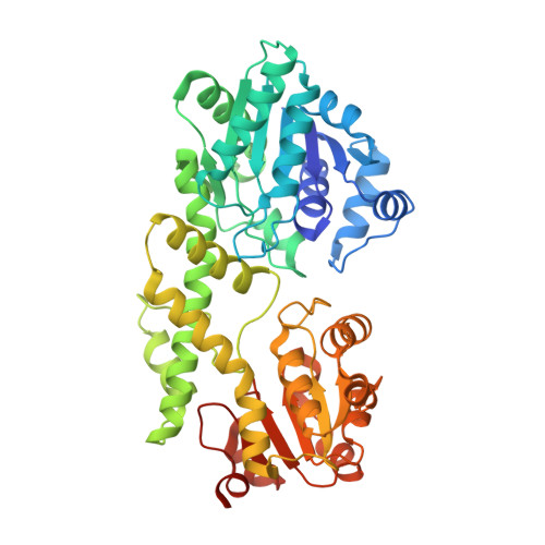
    Members of the Burkholderia cepacia complex (BCC) are serious respiratory pathogens in immunocompromised individuals and in patients with cystic fibrosis (CF). They are exceptionally resistant to many antimicrobial agents and have the capacity to spread between patients, leading to a decline in lung function and necrotizing pneumonia. BCC members often express a mucoid phenotype associated with the secretion of the exopolysaccharide (EPS) cepacian. There is much evidence supporting the fact that cepacian is a major virulence factor of BCC. UDP-glucose dehydrogenase (UGD) is responsible for the NAD-dependent 2-fold oxidation of UDP-glucose (UDP-Glc) to UDP-glucuronic acid (UDP-GlcA), which is a key step in cepacian biosynthesis. Here, we report the structure of BceC, determined at 1.75-Å resolution. Mutagenic studies were performed on the active sites of UGDs, and together with the crystallographic structures, they elucidate the molecular mechanism of this family of sugar nucleotide-modifying enzymes. Superposition with the structures of human and other bacterial UGDs showed an active site with high structural homology. This family contains a strictly conserved tyrosine residue (Y10 in BceC; shown in italics) within the glycine-rich motif (GXGYXG) of its N-terminal Rossmann-like domain. We constructed several BceC Y10 mutants, revealing only residual dehydrogenase activity and thus highlighting the importance of this conserved residue in the catalytic activity of BceC. Based on the literature of the UGD/GMD nucleotide sugar 6-dehydrogenase family and the kinetic and structural data we obtained for BceC, we determined Y10 as a key catalytic residue in a UGD rate-determining step, the final hydrolysis of the enzymatic thioester intermediate.


  • Organizational Affiliation

    Instituto de Tecnologia Química e Biológica, Universidade Nova de Lisboa, Apartado 127, P-2781-901 Oeiras, Portugal.


Macromolecules
Find similar proteins by:  (by identity cutoff)  |  3D Structure
Entity ID: 1
MoleculeChains Sequence LengthOrganismDetailsImage
UDP-GLUCOSE DEHYDROGENASE
A, B, C, D
478Burkholderia cepaciaMutation(s): 0 
EC: 1.1.1.22
UniProt
Find proteins for C9E261 (Burkholderia cepacia)
Explore C9E261 
Go to UniProtKB:  C9E261
Entity Groups  
Sequence Clusters30% Identity50% Identity70% Identity90% Identity95% Identity100% Identity
UniProt GroupC9E261
Sequence Annotations
Expand
  • Reference Sequence
Small Molecules
Ligands 4 Unique
IDChains Name / Formula / InChI Key2D Diagram3D Interactions
UGA
Query on UGA

Download Ideal Coordinates CCD File 
AA [auth D],
E [auth A],
M [auth B],
T [auth C]
URIDINE-5'-DIPHOSPHATE-GLUCURONIC ACID
C15 H22 N2 O18 P2
HDYANYHVCAPMJV-LXQIFKJMSA-N
SO4
Query on SO4

Download Ideal Coordinates CCD File 
BA [auth D]
CA [auth D]
DA [auth D]
F [auth A]
G [auth A]
BA [auth D],
CA [auth D],
DA [auth D],
F [auth A],
G [auth A],
H [auth A],
N [auth B],
O [auth B],
P [auth B],
Q [auth B],
R [auth B],
U [auth C],
V [auth C],
W [auth C]
SULFATE ION
O4 S
QAOWNCQODCNURD-UHFFFAOYSA-L
GOL
Query on GOL

Download Ideal Coordinates CCD File 
FA [auth D]
J [auth A]
K [auth A]
L [auth A]
Y [auth C]
FA [auth D],
J [auth A],
K [auth A],
L [auth A],
Y [auth C],
Z [auth C]
GLYCEROL
C3 H8 O3
PEDCQBHIVMGVHV-UHFFFAOYSA-N
ACT
Query on ACT

Download Ideal Coordinates CCD File 
EA [auth D],
I [auth A],
S [auth B],
X [auth C]
ACETATE ION
C2 H3 O2
QTBSBXVTEAMEQO-UHFFFAOYSA-M
Experimental Data & Validation

Experimental Data

  • Method: X-RAY DIFFRACTION
  • Resolution: 1.75 Å
  • R-Value Free: 0.197 
  • R-Value Work: 0.163 
  • R-Value Observed: 0.165 
  • Space Group: P 21 21 21
Unit Cell:
Length ( Å )Angle ( ˚ )
a = 97.631α = 90
b = 108.933β = 90
c = 187.708γ = 90
Software Package:
Software NamePurpose
PHENIXrefinement
MOSFLMdata reduction
SCALAdata scaling
PHASERphasing

Structure Validation

View Full Validation Report


Ligand Structure Quality Assessment 


Entry History 

Deposition Data

Revision History  (Full details and data files)

  • Version 1.0: 2011-07-27
    Type: Initial release
  • Version 1.1: 2019-03-06
    Changes: Data collection, Experimental preparation, Other
  • Version 1.2: 2019-05-08
    Changes: Data collection, Experimental preparation