2XFA

Crystal structure of Plasmodium berghei actin depolymerization factor 2


Experimental Data Snapshot

  • Method: X-RAY DIFFRACTION
  • Resolution: 2.10 Å
  • R-Value Free: 0.245 
  • R-Value Work: 0.186 
  • R-Value Observed: 0.190 

wwPDB Validation   3D Report Full Report


This is version 1.4 of the entry. See complete history


Literature

Crystal Structures Explain Functional Differences in the Two Actin Depolymerization Factors of the Malaria Parasite.

Singh, B.K.Sattler, J.M.Chatterjee, M.Huttu, J.Schuler, H.Kursula, I.

(2011) J Biol Chem 286: 28256

  • DOI: https://doi.org/10.1074/jbc.M111.211730
  • Primary Citation of Related Structures:  
    2XF1, 2XFA

  • PubMed Abstract: 

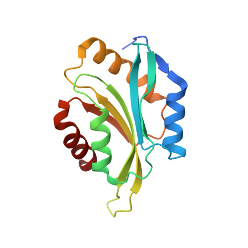
    Apicomplexan parasites, such as the malaria-causing Plasmodium, utilize an actin-based motor for motility and host cell invasion. The actin filaments of these parasites are unusually short, and actin polymerization is under strict control of a small set of regulatory proteins, which are poorly conserved with their mammalian orthologs. Actin depolymerization factors (ADFs) are among the most important actin regulators, affecting the rates of filament turnover in a multifaceted manner. Plasmodium has two ADFs that display low sequence homology with each other and with the higher eukaryotic family members. Here, we show that ADF2, like canonical ADF proteins but unlike ADF1, binds to both globular and filamentous actin, severing filaments and inducing nucleotide exchange on the actin monomer. The crystal structure of Plasmodium ADF1 shows major differences from the ADF consensus, explaining the lack of F-actin binding. Plasmodium ADF2 structurally resembles the canonical members of the ADF/cofilin family.


  • Organizational Affiliation

    Department of Biochemistry, University of Oulu, PO Box 3000, 90014 Oulu, Finland.


Macromolecules
Find similar proteins by:  (by identity cutoff)  |  3D Structure
Entity ID: 1
MoleculeChains Sequence LengthOrganismDetailsImage
ACTIN DEPOLYMERIZATION FACTOR 2
A, B
148Plasmodium bergheiMutation(s): 0 
UniProt
Find proteins for Q3YPH0 (Plasmodium berghei)
Explore Q3YPH0 
Go to UniProtKB:  Q3YPH0
Entity Groups  
Sequence Clusters30% Identity50% Identity70% Identity90% Identity95% Identity100% Identity
UniProt GroupQ3YPH0
Sequence Annotations
Expand
  • Reference Sequence
Experimental Data & Validation

Experimental Data

  • Method: X-RAY DIFFRACTION
  • Resolution: 2.10 Å
  • R-Value Free: 0.245 
  • R-Value Work: 0.186 
  • R-Value Observed: 0.190 
  • Space Group: P 21 21 2
Unit Cell:
Length ( Å )Angle ( ˚ )
a = 111.58α = 90
b = 57.9β = 90
c = 40.49γ = 90
Software Package:
Software NamePurpose
PHENIXrefinement
XDSdata reduction
XSCALEdata scaling
PHASERphasing

Structure Validation

View Full Validation Report


Entry History 

Deposition Data

Revision History  (Full details and data files)

  • Version 1.0: 2011-06-15
    Type: Initial release
  • Version 1.1: 2011-08-31
    Changes: Database references, Refinement description, Version format compliance
  • Version 1.2: 2018-01-17
    Changes: Data collection
  • Version 1.3: 2019-03-06
    Changes: Data collection, Experimental preparation
  • Version 1.4: 2023-12-20
    Changes: Data collection, Database references, Other, Refinement description