2XEJ

Human glutamate carboxypeptidase II in complex with ARM-M4, urea- based inhibitor


Experimental Data Snapshot

  • Method: X-RAY DIFFRACTION
  • Resolution: 1.78 Å
  • R-Value Free: 0.185 
  • R-Value Work: 0.157 
  • R-Value Observed: 0.157 

wwPDB Validation   3D Report Full Report


Ligand Structure Quality Assessment 


This is version 2.1 of the entry. See complete history


Literature

A Remote Arene-Binding Site on Prostate Specific Membrane Antigen Revealed by Antibody-Recruiting Small Molecules.

Zhang, A.X.Murelli, R.P.Barinka, C.Michel, J.Cocleaza, A.Jorgensen, W.L.Lubkowski, J.Spiegel, D.A.

(2010) J Am Chem Soc 132: 12711

  • DOI: https://doi.org/10.1021/ja104591m
  • Primary Citation of Related Structures:  
    2XEF, 2XEG, 2XEI, 2XEJ

  • PubMed Abstract: 

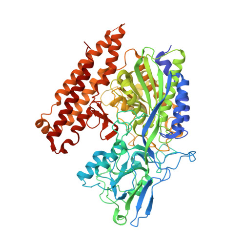
    Prostate specific membrane antigen (PSMA) is a membrane-bound glutamate carboxypeptidase overexpressed in many forms of prostate cancer. Our laboratory has recently disclosed a class of small molecules, called ARM-Ps (antibody-recruiting molecule targeting prostate cancer) that are capable of enhancing antibody-mediated immune recognition of prostate cancer cells. Interestingly, during the course of these studies, we found ARM-Ps to exhibit extraordinarily high potencies toward PSMA, compared to previously reported inhibitors. Here, we report in-depth biochemical, crystallographic, and computational investigations which elucidate the origin of the observed affinity enhancement. These studies reveal a previously unreported arene-binding site on PSMA, which we believe participates in an aromatic stacking interaction with ARMs. Although this site is composed of only a few amino acid residues, it drastically enhances small molecule binding affinity. These results provide critical insights into the design of PSMA-targeted small molecules for prostate cancer diagnosis and treatment; more broadly, the presence of similar arene-binding sites throughout the proteome could prove widely enabling in the optimization of small molecule-protein interactions.


  • Organizational Affiliation

    Department of Chemistry, Yale University, 225 Prospect Street, P.O. Box 208107, New Haven, Connecticut 06510-8107, USA.


Macromolecules
Find similar proteins by:  (by identity cutoff)  |  3D Structure
Entity ID: 1
MoleculeChains Sequence LengthOrganismDetailsImage
GLUTAMATE CARBOXYPEPTIDASE 2709Homo sapiensMutation(s): 0 
EC: 3.4.17.21
UniProt & NIH Common Fund Data Resources
Find proteins for Q04609 (Homo sapiens)
Explore Q04609 
Go to UniProtKB:  Q04609
PHAROS:  Q04609
GTEx:  ENSG00000086205 
Entity Groups  
Sequence Clusters30% Identity50% Identity70% Identity90% Identity95% Identity100% Identity
UniProt GroupQ04609
Sequence Annotations
Expand
  • Reference Sequence
Oligosaccharides

Help

Entity ID: 2
MoleculeChains Length2D Diagram Glycosylation3D Interactions
2-acetamido-2-deoxy-beta-D-glucopyranose-(1-4)-2-acetamido-2-deoxy-beta-D-glucopyranose
B, C, D
2N-Glycosylation
Glycosylation Resources
GlyTouCan:  G42666HT
GlyCosmos:  G42666HT
GlyGen:  G42666HT
Entity ID: 3
MoleculeChains Length2D Diagram Glycosylation3D Interactions
alpha-D-mannopyranose-(1-3)-beta-D-mannopyranose-(1-4)-2-acetamido-2-deoxy-beta-D-glucopyranose-(1-4)-2-acetamido-2-deoxy-beta-D-glucopyranose
E
4N-Glycosylation
Glycosylation Resources
GlyTouCan:  G81315DD
GlyCosmos:  G81315DD
GlyGen:  G81315DD
Small Molecules
Ligands 5 Unique
IDChains Name / Formula / InChI Key2D Diagram3D Interactions
OKD
Query on OKD

Download Ideal Coordinates CCD File 
M [auth A]N-({(1S)-1-CARBOXY-5-[4-(2,5,8,11,14-PENTAOXAPENTADEC-1-YL)-1H-1,2,3-TRIAZOL-1-YL]PENTYL}CARBAMOYL)-L-GLUTAMIC ACID
C24 H41 N5 O12
USQOBCXXBZGYAH-PMACEKPBSA-N
NAG
Query on NAG

Download Ideal Coordinates CCD File 
J [auth A],
K [auth A],
L [auth A]
2-acetamido-2-deoxy-beta-D-glucopyranose
C8 H15 N O6
OVRNDRQMDRJTHS-FMDGEEDCSA-N
ZN
Query on ZN

Download Ideal Coordinates CCD File 
F [auth A],
G [auth A]
ZINC ION
Zn
PTFCDOFLOPIGGS-UHFFFAOYSA-N
CA
Query on CA

Download Ideal Coordinates CCD File 
H [auth A]CALCIUM ION
Ca
BHPQYMZQTOCNFJ-UHFFFAOYSA-N
CL
Query on CL

Download Ideal Coordinates CCD File 
I [auth A]CHLORIDE ION
Cl
VEXZGXHMUGYJMC-UHFFFAOYSA-M
Binding Affinity Annotations 
IDSourceBinding Affinity
OKD Binding MOAD:  2XEJ Ki: 1.34 (nM) from 1 assay(s)
PDBBind:  2XEJ Ki: 1.34 (nM) from 1 assay(s)
Experimental Data & Validation

Experimental Data

  • Method: X-RAY DIFFRACTION
  • Resolution: 1.78 Å
  • R-Value Free: 0.185 
  • R-Value Work: 0.157 
  • R-Value Observed: 0.157 
  • Space Group: I 2 2 2
Unit Cell:
Length ( Å )Angle ( ˚ )
a = 101.508α = 90
b = 129.958β = 90
c = 159.246γ = 90
Software Package:
Software NamePurpose
REFMACrefinement
HKL-2000data reduction
HKL-2000data scaling
REFMACphasing

Structure Validation

View Full Validation Report


Ligand Structure Quality Assessment 


Entry History 

Deposition Data

Revision History  (Full details and data files)

  • Version 1.0: 2010-09-08
    Type: Initial release
  • Version 1.1: 2011-05-08
    Changes: Version format compliance
  • Version 1.2: 2011-07-13
    Changes: Version format compliance
  • Version 2.0: 2020-07-29
    Type: Remediation
    Reason: Carbohydrate remediation
    Changes: Advisory, Atomic model, Data collection, Derived calculations, Other, Structure summary
  • Version 2.1: 2023-12-20
    Changes: Advisory, Data collection, Database references, Refinement description, Structure summary