Three-Dimensional Solution NMR Structure of Apo-L75F-Trpr, a Temperature-Sensitive Mutant of the Tryptophan Repressor Protein.
Tyler, R., Pelczer, I., Carey, J., Copie, V.(2002) Biochemistry 41: 11954
- PubMed: 12356295
- DOI: https://doi.org/10.1021/bi020304t
- Primary Citation of Related Structures:
2XDI - PubMed Abstract:
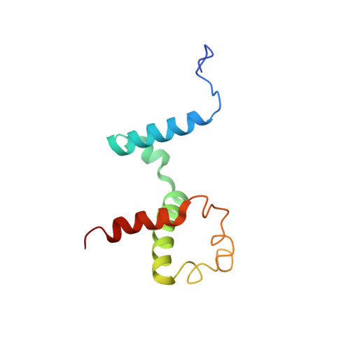
L75F-TrpR is a temperature-sensitive mutant of the tryptophan repressor protein of Escherichia coli in which surface-exposed residue leucine 75 in the DNA binding domain is replaced with phenylalanine. Biochemical and biophysical studies had suggested global alterations in dynamics for L75F-TrpR, although the structure was apparently similar to that of wild-type TrpR. Herein, we report the three-dimensional solution structure of apo-L75F-TrpR determined by multidimensional ((1)H, (15)N, and (13)C) solution NMR spectroscopy. An ensemble of structures was generated from 769 unique NOE-based distance restraints, 68 dihedral angle restraints, and 62 hydrogen bond distance restraints. Apo-L75F-TrpR exhibits a three-dimensional (3D) fold very similar to that of apo-WT-TrpR, with a dimeric core of four alpha-helices (A-C and F) from each subunit, and less well-defined D and E helical regions of the DNA binding domains. Despite their many similarities, wild-type and mutant proteins display significant chemical shift differences, one cluster of which is in the B-C turn, too distant to be ascribed solely to ring current effects from Phe75. Differences in NOE patterns and amide proton exchange rates are also observed in the B-C turn region. The data provide evidence that this point mutation exerts local effects on structure and stability in the DNA binding domain, and propagates long-range effects through the tertiary structure.
Organizational Affiliation:
Department of Chemistry and Biochemistry, Montana State University, 108 Gaines Hall, Bozeman, MT 59717, USA.