Structure of Bacteriophage T4 Endonuclease II Mutant E118A, a Tetrameric Giy-Yig Enzyme.
Andersson, C.E., Lagerback, P., Carlson, K.(2010) J Mol Biol 397: 1003
- PubMed: 20156453
- DOI: https://doi.org/10.1016/j.jmb.2010.01.076
- Primary Citation of Related Structures:
2WSH - PubMed Abstract:
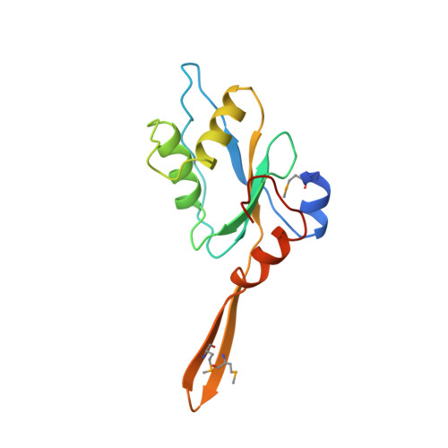
Coliphage T4 endonuclease II (EndoII), encoded by gene denA, is a small (16 kDa, 136 aa) enzyme belonging to the GIY-YIG family of endonucleases, which lacks a C-terminal domain corresponding to that providing most of the binding energy in the structurally characterized GIY-YIG endonucleases, I-TevI and UvrC. In vivo, it is involved in degradation of host DNA, permitting scavenging of host-derived nucleotides for phage DNA synthesis. EndoII primarily catalyzes single-stranded nicking of DNA; 5- to 10-fold less frequently double-stranded breaks are produced. The Glu118Ala mutant of EndoII was crystallized in space group P2(1) with four monomers in the asymmetric unit. The fold of the EndoII monomer is similar to that of the catalytic domains of UvrC and I-TevI. In contrast to these enzymes, EndoII forms a striking X-shaped tetrameric structure composed as a dimer of dimers, with a protruding hairpin domain not present in UvrC or I-TevI providing most of the dimerization and tetramerization interfaces. A bound phosphate ion in one of the four active sites of EndoII likely mimics the scissile phosphate in a true substrate complex. In silico docking experiments showed that a protruding loop containing a nuclease-associated modular domain 3 element is likely to be involved in substrate binding, as well as residues forming a separate nucleic acid binding surface adjacent to the active site. The positioning of these sites within the EndoII primary dimer suggests that the substrate would bind to a primary EndoII dimer diagonally over the active sites, requiring significant distortion of the enzyme or the substrate DNA, or both, for simultaneous nicking of both DNA strands. The scarcity of potential nucleic acid binding residues between the active sites indicates that EndoII may bind its substrate inefficiently across the two sites in the dimer, offering a plausible explanation for the catalytic preponderance of single-strand nicks. Mutations analyzed in earlier functional studies are discussed in their structural context.
Organizational Affiliation:
Department of Molecular Biology, Swedish University of Agricultural Sciences, Uppsala, Sweden. evalena@xray.bmc.uu.se