A Genetically Encoded Photoactivatable Rac Controls the Motility of Living Cells.
Wu, Y.I., Frey, D., Lungu, O.I., Jaehrig, A., Schlichting, I., Kuhlman, B., Hahn, K.M.(2009) Nature 461: 104
- PubMed: 19693014
- DOI: https://doi.org/10.1038/nature08241
- Primary Citation of Related Structures:
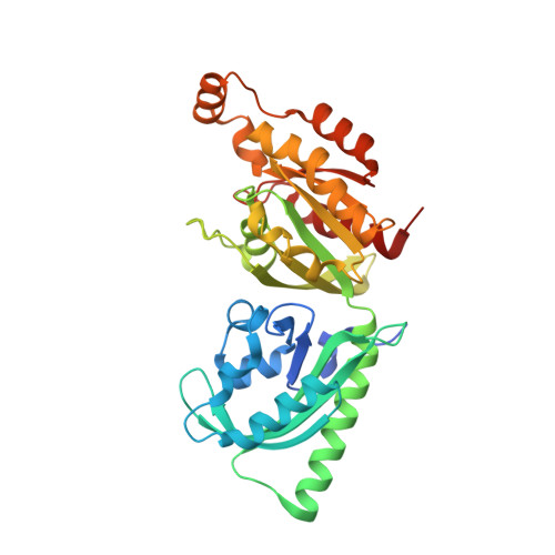
2WKP, 2WKQ, 2WKR - PubMed Abstract:
The precise spatio-temporal dynamics of protein activity are often critical in determining cell behaviour, yet for most proteins they remain poorly understood; it remains difficult to manipulate protein activity at precise times and places within living cells. Protein activity has been controlled by light, through protein derivatization with photocleavable moieties or using photoreactive small-molecule ligands. However, this requires use of toxic ultraviolet wavelengths, activation is irreversible, and/or cell loading is accomplished via disruption of the cell membrane (for example, through microinjection). Here we have developed a new approach to produce genetically encoded photoactivatable derivatives of Rac1, a key GTPase regulating actin cytoskeletal dynamics in metazoan cells. Rac1 mutants were fused to the photoreactive LOV (light oxygen voltage) domain from phototropin, sterically blocking Rac1 interactions until irradiation unwound a helix linking LOV to Rac1. Photoactivatable Rac1 (PA-Rac1) could be reversibly and repeatedly activated using 458- or 473-nm light to generate precisely localized cell protrusions and ruffling. Localized Rac activation or inactivation was sufficient to produce cell motility and control the direction of cell movement. Myosin was involved in Rac control of directionality but not in Rac-induced protrusion, whereas PAK was required for Rac-induced protrusion. PA-Rac1 was used to elucidate Rac regulation of RhoA in cell motility. Rac and Rho coordinate cytoskeletal behaviours with seconds and submicrometre precision. Their mutual regulation remains controversial, with data indicating that Rac inhibits and/or activates Rho. Rac was shown to inhibit RhoA in mouse embryonic fibroblasts, with inhibition modulated at protrusions and ruffles. A PA-Rac crystal structure and modelling revealed LOV-Rac interactions that will facilitate extension of this photoactivation approach to other proteins.
Organizational Affiliation:
Department of Pharmacology, University of North Carolina, Chapel Hill, North Carolina 27599, USA. yiwu@med.unc.edu