2WJI

Structure and function of the FeoB G-domain from Methanococcus jannaschii


Experimental Data Snapshot

  • Method: X-RAY DIFFRACTION
  • Resolution: 1.90 Å
  • R-Value Free: 0.269 
  • R-Value Work: 0.218 
  • R-Value Observed: 0.221 

wwPDB Validation   3D Report Full Report


Ligand Structure Quality Assessment 


This is version 1.3 of the entry. See complete history


Literature

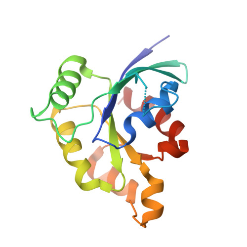
Structure and Function of the Feob G-Domain from Methanococcus Jannaschii

Koester, S.Wehner, M.Herrmann, C.Kuehlbrandt, W.Yildiz, O.

(2009) J Mol Biol 392: 405

  • DOI: https://doi.org/10.1016/j.jmb.2009.07.020
  • Primary Citation of Related Structures:  
    2WJG, 2WJH, 2WJI, 2WJJ

  • PubMed Abstract: 

    FeoB in bacteria and archaea is involved in the uptake of ferrous iron (Fe(2+)), an important cofactor in biological electron transfer and catalysis. Unlike any other known prokaryotic membrane protein, FeoB contains a GTP-binding domain at its N-terminus. We determined high-resolution X-ray structures of the FeoB G-domain from Methanococcus jannaschii with and without bound GDP or Mg(2+)-GppNHp. The G-domain forms the same dimer in all three structures, with the nucleotide-binding pockets at the dimer interface, as in the ATP-binding domain of ABC transporters. The G-domain follows the typical fold of nucleotide-binding proteins, with a beta-strand inserted in switch I that becomes partially disordered upon GTP binding. Switch II does not contact the nucleotide directly and does not change its conformation in response to the bound nucleotide. Release of the nucleotide causes a rearrangement of loop L6, which we identified as the G5 region of FeoB. Together with the C-terminal helix, this loop may transmit the information about the nucleotide-bound state from the G-domain to the transmembrane region of FeoB.


  • Organizational Affiliation

    Department of Structural Biology, Max Planck Institute of Biophysics, Frankfurt am Main, Germany.


Macromolecules
Find similar proteins by:  (by identity cutoff)  |  3D Structure
Entity ID: 1
MoleculeChains Sequence LengthOrganismDetailsImage
FERROUS IRON TRANSPORT PROTEIN B HOMOLOG
A, B
165Methanocaldococcus jannaschiiMutation(s): 0 
UniProt
Find proteins for Q57986 (Methanocaldococcus jannaschii (strain ATCC 43067 / DSM 2661 / JAL-1 / JCM 10045 / NBRC 100440))
Explore Q57986 
Go to UniProtKB:  Q57986
Entity Groups  
Sequence Clusters30% Identity50% Identity70% Identity90% Identity95% Identity100% Identity
UniProt GroupQ57986
Sequence Annotations
Expand
  • Reference Sequence
Experimental Data & Validation

Experimental Data

  • Method: X-RAY DIFFRACTION
  • Resolution: 1.90 Å
  • R-Value Free: 0.269 
  • R-Value Work: 0.218 
  • R-Value Observed: 0.221 
  • Space Group: P 1 21 1
Unit Cell:
Length ( Å )Angle ( ˚ )
a = 49.55α = 90
b = 68.45β = 96.97
c = 52.99γ = 90
Software Package:
Software NamePurpose
PHENIXrefinement
XDSdata reduction
XSCALEdata scaling
PHASERphasing

Structure Validation

View Full Validation Report


Ligand Structure Quality Assessment 


Entry History 

Deposition Data

Revision History  (Full details and data files)

  • Version 1.0: 2009-07-28
    Type: Initial release
  • Version 1.1: 2011-05-19
    Changes: Version format compliance
  • Version 1.2: 2011-07-13
    Changes: Version format compliance
  • Version 1.3: 2023-12-13
    Changes: Data collection, Database references, Derived calculations, Other, Refinement description