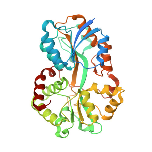
Futa2 is a Ferric Binding Protein from Synechocystis Pcc 6803.
Badarau, A., Firbank, S.J., Waldron, K.J., Yanagisawa, S., Robinson, N.J., Banfield, M.J., Dennison, C.(2008) J Biol Chem 283: 12520
- PubMed: 18252722
- DOI: https://doi.org/10.1074/jbc.M709907200
- Primary Citation of Related Structures:
2VOZ, 2VP1 - PubMed Abstract:
Synechocystis PCC 6803 has a high demand for iron (10 times greater than Escherichia coli) to sustain photosynthesis and is unusual in possessing at least two putative iron-binding proteins of a type normally associated with ATP-binding cassette-type importers. It has been suggested that one of these, FutA2, binds ferrous iron, but herein we clearly demonstrate that this protein avidly binds Fe(III), the oxidation state preference of periplasmic iron-binding proteins. Structures of apo-FutA2 and Fe-FutA2 have been determined at 1.7 and 2.7A, respectively. The metal ion is bound in a distorted trigonal bipyramidal arrangement with no exogenous anions as ligands. The metal-binding environment, including the second coordination sphere and charge properties, is consistent with a preference for Fe(III). Atypically, FutA2 has a Tat signal peptide, and its inability to coordinate divalent cations may be crucial to prevent metals from binding to the folded protein prior to export from the cytosol. A loop containing the His(43) ligand undergoes considerable movement in apo-versus Fe-FutA2 and may control metal release to the importer. Although these data are consistent with FutA2 being the periplasmic component involved in iron uptake, deletion of another putative ferric binding protein, FutA1, has a greater effect on the accumulation of iron and is more analogous to a DeltafutA1DeltafutA2 double mutant than DeltafutA2. Here, we also discover that there is a reduced level of ferric FutA2 in the periplasm of the DeltafutA1 mutant providing an explanation for its severe iron-uptake phenotype.
Organizational Affiliation:
Institute for Cell and Molecular Biosciences, Medical School, Newcastle University, Newcastle upon Tyne NE2 4HH, United Kingdom.