2VMD

Structure of the complex of discoidin II from Dictyostelium discoideum with beta-methyl-galactose


Experimental Data Snapshot

  • Method: X-RAY DIFFRACTION
  • Resolution: 1.90 Å
  • R-Value Free: 0.227 
  • R-Value Work: 0.178 
  • R-Value Observed: 0.181 

wwPDB Validation   3D Report Full Report


Ligand Structure Quality Assessment 


This is version 1.4 of the entry. See complete history


Literature

Structure Determination of Discoidin II from Dictyostelium Discoideum and Carbohydrate Binding Properties of the Lectin Domain.

Aragao, K.S.Satre, M.Imberty, A.Varrot, A.

(2008) Proteins 73: 43

  • DOI: https://doi.org/10.1002/prot.22038
  • Primary Citation of Related Structures:  
    2VM9, 2VMC, 2VMD, 2VME

  • PubMed Abstract: 

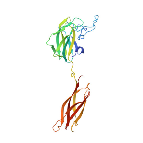
    The social amoeba Dictyostelium discoideum adopts a cohesive stage upon starvation and then produces Discoidin I and II, two proteins able to bind galactose and N-acetyl-galactosamine. The N-terminal domain or discoidin domain (DS) is widely distributed in eukaryotes where it plays a role in extracellular matrix binding while the C-terminal domain displays sequence similarities to invertebrate lectins. We present the first X-ray structures of the wild-type and recombinant Discoidin II in unliganded state and in complex with monosaccharides. The protein forms a homotrimer which presents two binding surfaces situated on the opposite boundaries of the structure. The binding sites of the N-terminal domain contain PEG molecules that could mimics binding of natural ligand. The C-terminal lectin domain interactions with N-acetyl-D-galactosamine and methyl-beta-galactoside are described. The carbohydrate binding sites are located at the interface between monomers. Specificity for galacto configuration can be rationalized since the axial O4 hydroxyl group is involved in several hydrogen bonds with protein side chains. Titration microcalorimetry allowed characterization of affinity and demonstrated the enthalpy-driven character of the interaction. Those results highlight the structural differentiation of the DS domain involved in many cell-adhesion processes from the lectin activity of Dictyostelium discoidins.


  • Organizational Affiliation

    CERMAV-CNRS (affiliated with University Joseph Fourier), Grenoble, France.


Macromolecules
Find similar proteins by:  (by identity cutoff)  |  3D Structure
Entity ID: 1
MoleculeChains Sequence LengthOrganismDetailsImage
DISCOIDIN-2257Dictyostelium discoideum AX2Mutation(s): 0 
UniProt
Find proteins for P42530 (Dictyostelium discoideum)
Explore P42530 
Go to UniProtKB:  P42530
Entity Groups  
Sequence Clusters30% Identity50% Identity70% Identity90% Identity95% Identity100% Identity
UniProt GroupP42530
Sequence Annotations
Expand
  • Reference Sequence
Small Molecules
Ligands 5 Unique
IDChains Name / Formula / InChI Key2D Diagram3D Interactions
1PG
Query on 1PG

Download Ideal Coordinates CCD File 
E [auth A],
F [auth A]
2-(2-{2-[2-(2-METHOXY-ETHOXY)-ETHOXY]-ETHOXY}-ETHOXY)-ETHANOL
C11 H24 O6
SLNYBUIEAMRFSZ-UHFFFAOYSA-N
MBG
Query on MBG

Download Ideal Coordinates CCD File 
D [auth A]methyl beta-D-galactopyranoside
C7 H14 O6
HOVAGTYPODGVJG-VOQCIKJUSA-N
EDO
Query on EDO

Download Ideal Coordinates CCD File 
G [auth A]1,2-ETHANEDIOL
C2 H6 O2
LYCAIKOWRPUZTN-UHFFFAOYSA-N
CA
Query on CA

Download Ideal Coordinates CCD File 
C [auth A]CALCIUM ION
Ca
BHPQYMZQTOCNFJ-UHFFFAOYSA-N
CL
Query on CL

Download Ideal Coordinates CCD File 
B [auth A]CHLORIDE ION
Cl
VEXZGXHMUGYJMC-UHFFFAOYSA-M
Binding Affinity Annotations 
IDSourceBinding Affinity
MBG Binding MOAD:  2VMD Kd: 9.50e+5 (nM) from 1 assay(s)
PDBBind:  2VMD Kd: 9.50e+5 (nM) from 1 assay(s)
Experimental Data & Validation

Experimental Data

  • Method: X-RAY DIFFRACTION
  • Resolution: 1.90 Å
  • R-Value Free: 0.227 
  • R-Value Work: 0.178 
  • R-Value Observed: 0.181 
  • Space Group: H 3 2
Unit Cell:
Length ( Å )Angle ( ˚ )
a = 81.14α = 90
b = 81.14β = 90
c = 259.28γ = 120
Software Package:
Software NamePurpose
REFMACrefinement
XDSdata reduction
SCALAdata scaling
AMoREphasing

Structure Validation

View Full Validation Report


Ligand Structure Quality Assessment 


Entry History 

Deposition Data

Revision History  (Full details and data files)

  • Version 1.0: 2008-07-01
    Type: Initial release
  • Version 1.1: 2011-06-02
    Changes: Version format compliance
  • Version 1.2: 2011-07-13
    Changes: Version format compliance
  • Version 1.3: 2020-07-29
    Type: Remediation
    Reason: Carbohydrate remediation
    Changes: Data collection, Derived calculations, Other, Structure summary
  • Version 1.4: 2023-12-13
    Changes: Data collection, Database references, Refinement description, Structure summary