2VGK

Crystal structure of Actinomadura R39 DD-peptidase complexed with a peptidoglycan-mimetic cephalosporin


Experimental Data Snapshot

  • Method: X-RAY DIFFRACTION
  • Resolution: 2.25 Å
  • R-Value Free: 0.286 
  • R-Value Work: 0.230 
  • R-Value Observed: 0.233 

wwPDB Validation   3D Report Full Report


Ligand Structure Quality Assessment 


This is version 1.3 of the entry. See complete history


Literature

Crystal Structures of Complexes of Bacterial Dd-Peptidases with Peptidoglycan-Mimetic Ligands: The Substrate Specificity Puzzle.

Sauvage, E.Powell, A.J.Heilemann, J.Josephin, H.R.Charlier, P.Davies, C.Pratt, R.F.

(2008) J Mol Biol 381: 383

  • DOI: https://doi.org/10.1016/j.jmb.2008.06.012
  • Primary Citation of Related Structures:  
    2VGJ, 2VGK, 3BEB, 3BEC

  • PubMed Abstract: 

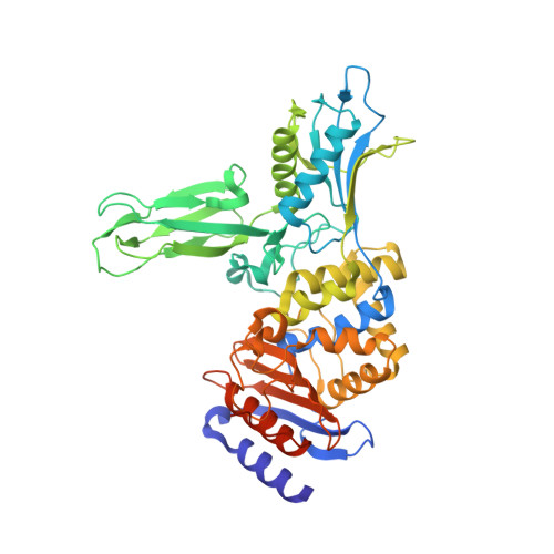
    The X-ray crystal structures of covalent complexes of the Actinomadura R39 dd-peptidase and Escherichia coli penicillin-binding protein (PBP) 5 with beta-lactams bearing peptidoglycan-mimetic side chains have been determined. The structure of the hydrolysis product of an analogous peptide bound noncovalently to the former enzyme has also been obtained. The R39 DD-peptidase structures reveal the presence of a specific binding site for the D-alpha-aminopimelyl side chain, characteristic of the stem peptide of Actinomadura R39. This binding site features a hydrophobic cleft for the pimelyl methylene groups and strong hydrogen bonding to the polar terminus. Both of these active site elements are provided by amino acid side chains from two separate domains of the protein. In contrast, no clear electron density corresponding to the terminus of the peptidoglycan-mimetic side chains is present when these beta-lactams are covalently bound to PBP5. There is, therefore, no indication of a specific side-chain binding site in this enzyme. These results are in agreement with those from kinetics studies published earlier and support the general prediction made at the time of a direct correlation between kinetics and structural evidence. The essential high-molecular-mass PBPs have demonstrated, to date, no specific reactivity with peptidoglycan-mimetic peptide substrates and beta-lactam inhibitors and, thus, probably do not possess a specific substrate-binding site of the type demonstrated here with the R39 DD-peptidase. This striking deficiency may represent a sophisticated defense mechanism against low-molecular-mass substrate-analogue inhibitors/antibiotics; its discovery should focus new inhibitor design.


  • Organizational Affiliation

    Centre d'Ingéniere des Proteines, Université de Liège, B-4000 Sart Tilman, Liège, Belgium.


Macromolecules
Find similar proteins by:  (by identity cutoff)  |  3D Structure
Entity ID: 1
MoleculeChains Sequence LengthOrganismDetailsImage
D-ALANYL-D-ALANINE CARBOXYPEPTIDASE
A, B, C, D
489Actinomadura sp. R39Mutation(s): 0 
EC: 3.4.16.4
UniProt
Find proteins for P39045 (Actinomadura sp. (strain R39))
Explore P39045 
Go to UniProtKB:  P39045
Entity Groups  
Sequence Clusters30% Identity50% Identity70% Identity90% Identity95% Identity100% Identity
UniProt GroupP39045
Sequence Annotations
Expand
  • Reference Sequence
Small Molecules
Ligands 3 Unique
IDChains Name / Formula / InChI Key2D Diagram3D Interactions
REZ
Query on REZ

Download Ideal Coordinates CCD File 
E [auth A],
M [auth B],
S [auth C]
(2R)-2-AMINO-7-{[(1R)-1-CARBOXYETHYL]AMINO}-7-OXOHEPTANOIC ACID
C10 H18 N2 O5
GMHVWLJCMCGBKZ-RNFRBKRXSA-N
SO4
Query on SO4

Download Ideal Coordinates CCD File 
AA [auth D]
BA [auth D]
CA [auth D]
F [auth A]
G [auth A]
AA [auth D],
BA [auth D],
CA [auth D],
F [auth A],
G [auth A],
H [auth A],
I [auth A],
J [auth A],
N [auth B],
O [auth B],
P [auth B],
Q [auth B],
R [auth B],
T [auth C],
U [auth C],
V [auth C],
W [auth C],
X [auth C],
Y [auth D],
Z [auth D]
SULFATE ION
O4 S
QAOWNCQODCNURD-UHFFFAOYSA-L
MG
Query on MG

Download Ideal Coordinates CCD File 
DA [auth D],
EA [auth D],
K [auth A],
L [auth A]
MAGNESIUM ION
Mg
JLVVSXFLKOJNIY-UHFFFAOYSA-N
Experimental Data & Validation

Experimental Data

  • Method: X-RAY DIFFRACTION
  • Resolution: 2.25 Å
  • R-Value Free: 0.286 
  • R-Value Work: 0.230 
  • R-Value Observed: 0.233 
  • Space Group: P 1 21 1
Unit Cell:
Length ( Å )Angle ( ˚ )
a = 105.14α = 90
b = 93.35β = 94.98
c = 109.1γ = 90
Software Package:
Software NamePurpose
REFMACrefinement
XDSdata reduction
SCALAdata scaling

Structure Validation

View Full Validation Report


Ligand Structure Quality Assessment 


Entry History 

Deposition Data

Revision History  (Full details and data files)

  • Version 1.0: 2008-11-25
    Type: Initial release
  • Version 1.1: 2011-05-08
    Changes: Version format compliance
  • Version 1.2: 2011-07-13
    Changes: Version format compliance
  • Version 1.3: 2023-12-13
    Changes: Data collection, Database references, Derived calculations, Other, Refinement description