2V8Q

Crystal structure of the regulatory fragment of mammalian AMPK in complexes with AMP


Experimental Data Snapshot

  • Method: X-RAY DIFFRACTION
  • Resolution: 2.10 Å
  • R-Value Free: 0.237 
  • R-Value Work: 0.212 
  • R-Value Observed: 0.213 

wwPDB Validation   3D Report Full Report


Ligand Structure Quality Assessment 


This is version 1.1 of the entry. See complete history


Literature

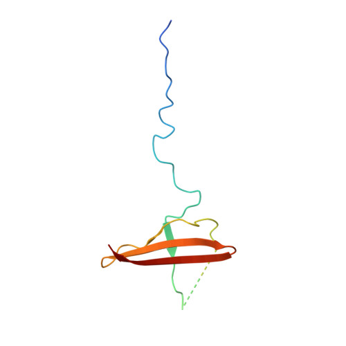
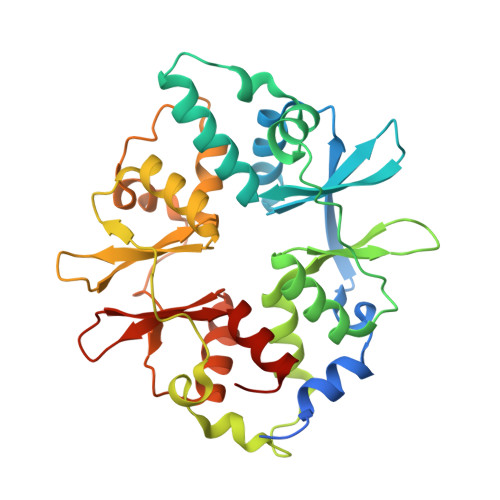
Structural Basis for AMP Binding to Mammalian AMP-Activated Protein Kinase

Xiao, B.Heath, R.Saiu, P.Leiper, F.C.Leone, P.Jing, C.Walker, P.A.Haire, L.Eccleston, J.F.Davis, C.T.Martin, S.R.Carling, D.Gamblin, S.J.

(2007) Nature 449: 496

  • DOI: https://doi.org/10.1038/nature06161
  • Primary Citation of Related Structures:  
    2V8Q, 2V92, 2V9J

  • PubMed Abstract: 

    AMP-activated protein kinase (AMPK) regulates cellular metabolism in response to the availability of energy and is therefore a target for type II diabetes treatment. It senses changes in the ratio of AMP/ATP by binding both species in a competitive manner. Thus, increases in the concentration of AMP activate AMPK resulting in the phosphorylation and differential regulation of a series of downstream targets that control anabolic and catabolic pathways. We report here the crystal structure of the regulatory fragment of mammalian AMPK in complexes with AMP and ATP. The phosphate groups of AMP/ATP lie in a groove on the surface of the gamma domain, which is lined with basic residues, many of which are associated with disease-causing mutations. Structural and solution studies reveal that two sites on the gamma domain bind either AMP or Mg.ATP, whereas a third site contains a tightly bound AMP that does not exchange. Our binding studies indicate that under physiological conditions AMPK mainly exists in its inactive form in complex with Mg.ATP, which is much more abundant than AMP. Our modelling studies suggest how changes in the concentration of AMP ([AMP]) enhance AMPK activity levels. The structure also suggests a mechanism for propagating AMP/ATP signalling whereby a phosphorylated residue from the alpha and/or beta subunits binds to the gamma subunit in the presence of AMP but not when ATP is bound.


  • Organizational Affiliation

    MRC National Institute for Medical Research, The Ridgeway, Mill Hill, London NW7 1AA, UK.


Macromolecules
Find similar proteins by:  (by identity cutoff)  |  3D Structure
Entity ID: 1
MoleculeChains Sequence LengthOrganismDetailsImage
5'-AMP-ACTIVATED PROTEIN KINASE CATALYTIC SUBUNIT ALPHA-1157Rattus norvegicusMutation(s): 0 
EC: 2.7.11.1
UniProt
Find proteins for P54645 (Rattus norvegicus)
Explore P54645 
Go to UniProtKB:  P54645
Entity Groups  
Sequence Clusters30% Identity50% Identity70% Identity90% Identity95% Identity100% Identity
UniProt GroupP54645
Sequence Annotations
Expand
  • Reference Sequence
Find similar proteins by:  (by identity cutoff)  |  3D Structure
Entity ID: 2
MoleculeChains Sequence LengthOrganismDetailsImage
5'-AMP-ACTIVATED PROTEIN KINASE SUBUNIT BETA-287Homo sapiensMutation(s): 0 
UniProt & NIH Common Fund Data Resources
Find proteins for O43741 (Homo sapiens)
Explore O43741 
Go to UniProtKB:  O43741
PHAROS:  O43741
GTEx:  ENSG00000131791 
Entity Groups  
Sequence Clusters30% Identity50% Identity70% Identity90% Identity95% Identity100% Identity
UniProt GroupO43741
Sequence Annotations
Expand
  • Reference Sequence
Find similar proteins by:  (by identity cutoff)  |  3D Structure
Entity ID: 3
MoleculeChains Sequence LengthOrganismDetailsImage
5'-AMP-ACTIVATED PROTEIN KINASE SUBUNIT GAMMA-1C [auth E]330Rattus norvegicusMutation(s): 0 
UniProt
Find proteins for P80385 (Rattus norvegicus)
Explore P80385 
Go to UniProtKB:  P80385
Entity Groups  
Sequence Clusters30% Identity50% Identity70% Identity90% Identity95% Identity100% Identity
UniProt GroupP80385
Sequence Annotations
Expand
  • Reference Sequence
Small Molecules
Binding Affinity Annotations 
IDSourceBinding Affinity
AMP Binding MOAD:  2V8Q Kd: 2.45e+4 (nM) from 1 assay(s)
BindingDB:  2V8Q Kd: 3700 (nM) from 1 assay(s)
EC50: 1519 (nM) from 1 assay(s)
Experimental Data & Validation

Experimental Data

  • Method: X-RAY DIFFRACTION
  • Resolution: 2.10 Å
  • R-Value Free: 0.237 
  • R-Value Work: 0.212 
  • R-Value Observed: 0.213 
  • Space Group: P 21 21 21
Unit Cell:
Length ( Å )Angle ( ˚ )
a = 48.522α = 90
b = 119.39β = 90
c = 129.31γ = 90
Software Package:
Software NamePurpose
REFMACrefinement

Structure Validation

View Full Validation Report


Ligand Structure Quality Assessment 


Entry History 

Revision History  (Full details and data files)

  • Version 1.0: 2007-09-25
    Type: Initial release
  • Version 1.1: 2011-07-13
    Changes: Advisory, Version format compliance