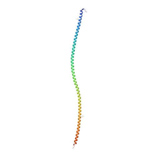
2V71

Coiled-coil region of NudEL


Experimental Data Snapshot

  • Method: X-RAY DIFFRACTION
  • Resolution: 2.24 Å
  • R-Value Free: 0.290 
  • R-Value Work: 0.253 
  • R-Value Observed: 0.255 

wwPDB Validation   3D Report Full Report


This is version 1.2 of the entry. See complete history


Literature

The Structure of the Coiled-Coil Domain of Ndel1 and the Basis of its Interaction with Lis1, the Causal Protein of Miller-Dieker Lissencephaly.

Derewenda, U.Tarricone, C.Choi, W.C.Cooper, D.R.Lukasik, S.Perrina, F.Tripathy, A.Kim, M.H.Cafiso, D.S.Musacchio, A.Derewenda, Z.S.

(2007) Structure 15: 1467

  • DOI: https://doi.org/10.1016/j.str.2007.09.015
  • Primary Citation of Related Structures:  
    2V66, 2V71

  • PubMed Abstract: 

    Ndel1 and Nde1 are homologous and evolutionarily conserved proteins, with critical roles in cell division, neuronal migration, and other physiological phenomena. These functions are dependent on their interactions with the retrograde microtubule motor dynein and with its regulator Lis1--a product of the causal gene for isolated lissencephaly sequence (ILS) and Miller-Dieker lissencephaly. The molecular basis of the interactions of Ndel1 and Nde1 with Lis1 is not known. Here, we present a crystallographic study of two fragments of the coiled-coil domain of Ndel1, one of which reveals contiguous high-quality electron density for residues 10-166, the longest such structure reported by X-ray diffraction at high resolution. Together with complementary solution studies, our structures reveal how the Ndel1 coiled coil forms a stable parallel homodimer and suggest mechanisms by which the Lis1-interacting domain can be regulated to maintain a conformation in which two supercoiled alpha helices cooperatively bind to a Lis1 homodimer.


  • Organizational Affiliation

    Department of Molecular Physiology and Biological Physics, University of Virginia, Charlottesville, VA 22908, USA.


Macromolecules
Find similar proteins by:  (by identity cutoff)  |  3D Structure
Entity ID: 1
MoleculeChains Sequence LengthOrganismDetailsImage
NUCLEAR DISTRIBUTION PROTEIN NUDE-LIKE 1
A, B
189Rattus norvegicusMutation(s): 2 
UniProt
Find proteins for Q78PB6 (Rattus norvegicus)
Explore Q78PB6 
Go to UniProtKB:  Q78PB6
Entity Groups  
Sequence Clusters30% Identity50% Identity70% Identity90% Identity95% Identity100% Identity
UniProt GroupQ78PB6
Sequence Annotations
Expand
  • Reference Sequence
Small Molecules
Modified Residues  1 Unique
IDChains TypeFormula2D DiagramParent
MSE
Query on MSE
A, B
L-PEPTIDE LINKINGC5 H11 N O2 SeMET
Experimental Data & Validation

Experimental Data

  • Method: X-RAY DIFFRACTION
  • Resolution: 2.24 Å
  • R-Value Free: 0.290 
  • R-Value Work: 0.253 
  • R-Value Observed: 0.255 
  • Space Group: C 1 2 1
Unit Cell:
Length ( Å )Angle ( ˚ )
a = 89.872α = 90
b = 44.975β = 97.35
c = 124.377γ = 90
Software Package:
Software NamePurpose
REFMACrefinement
HKL-2000data reduction
HKL-2000data scaling
SOLVEphasing

Structure Validation

View Full Validation Report


Entry History 

Deposition Data

Revision History  (Full details and data files)

  • Version 1.0: 2007-11-27
    Type: Initial release
  • Version 1.1: 2011-07-13
    Changes: Advisory, Version format compliance
  • Version 1.2: 2017-06-28
    Changes: Data collection