2V0I

Characterization of Substrate Binding and Catalysis of the Potential Antibacterial Target N-acetylglucosamine-1-phosphate Uridyltransferase (GlmU)


Experimental Data Snapshot

  • Method: X-RAY DIFFRACTION
  • Resolution: 1.89 Å
  • R-Value Free: 0.210 
  • R-Value Work: 0.193 
  • R-Value Observed: 0.194 

wwPDB Validation   3D Report Full Report


Ligand Structure Quality Assessment 


This is version 1.2 of the entry. See complete history


Literature

Characterization of Substrate Binding and Catalysis in the Potential Antibacterial Target N-Acetylglucosamine-1-Phosphate Uridyltransferase (Glmu).

Mochalkin, I.Lightle, S.Zhu, Y.Ohren, J.F.Spessard, C.Chirgadze, N.Y.Banotai, C.Melnick, M.Mcdowell, L.

(2007) Protein Sci 16: 2657

  • DOI: https://doi.org/10.1110/ps.073135107
  • Primary Citation of Related Structures:  
    2V0H, 2V0I, 2V0J, 2V0K, 2V0L

  • PubMed Abstract: 

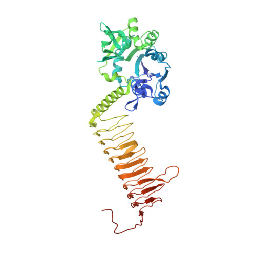
    N-Acetylglucosamine-1-phosphate uridyltransferase (GlmU) catalyzes the first step in peptidoglycan biosynthesis in both Gram-positive and Gram-negative bacteria. The products of the GlmU reaction are essential for bacterial survival, making this enzyme an attractive target for antibiotic drug discovery. A series of Haemophilus influenzae GlmU (hiGlmU) structures were determined by X-ray crystallography in order to provide structural and functional insights into GlmU activity and inhibition. The information derived from these structures was combined with biochemical characterization of the K25A, Q76A, D105A, Y103A, V223A, and E224A hiGlmU mutants in order to map these active-site residues to catalytic activity of the enzyme and refine the mechanistic model of the GlmU uridyltransferase reaction. These studies suggest that GlmU activity follows a sequential substrate-binding order that begins with UTP binding noncovalently to the GlmU enzyme. The uridyltransferase active site then remains in an open apo-like conformation until N-acetylglucosamine-1-phosphate (GlcNAc-1-P) binds and induces a conformational change at the GlcNAc-binding subsite. Following the binding of GlcNAc-1-P to the UTP-charged uridyltransferase active site, the non-esterified oxygen of GlcNAc-1-P performs a nucleophilic attack on the alpha-phosphate group of UTP. The new data strongly suggest that the mechanism of phosphotransfer in the uridyltransferase reaction in GlmU is primarily through an associative mechanism with a pentavalent phosphate intermediate and an inversion of stereochemistry. Finally, the structural and biochemical characterization of the uridyltransferase active site and catalytic mechanism described herein provides a basis for the structure-guided design of novel antibacterial agents targeting GlmU activity.


  • Organizational Affiliation

    Pfizer Global Research and Development, Michigan Laboratories, Ann Arbor, Michigan 48105, USA. Igor.Mochalkin@pfizer.com


Macromolecules
Find similar proteins by:  (by identity cutoff)  |  3D Structure
Entity ID: 1
MoleculeChains Sequence LengthOrganismDetailsImage
BIFUNCTIONAL PROTEIN GLMU456Haemophilus influenzaeMutation(s): 0 
EC: 2
UniProt
Find proteins for P43889 (Haemophilus influenzae (strain ATCC 51907 / DSM 11121 / KW20 / Rd))
Explore P43889 
Go to UniProtKB:  P43889
Entity Groups  
Sequence Clusters30% Identity50% Identity70% Identity90% Identity95% Identity100% Identity
UniProt GroupP43889
Sequence Annotations
Expand
  • Reference Sequence
Small Molecules
Ligands 4 Unique
IDChains Name / Formula / InChI Key2D Diagram3D Interactions
UD1
Query on UD1

Download Ideal Coordinates CCD File 
B [auth A]URIDINE-DIPHOSPHATE-N-ACETYLGLUCOSAMINE
C17 H27 N3 O17 P2
LFTYTUAZOPRMMI-CFRASDGPSA-N
PG4
Query on PG4

Download Ideal Coordinates CCD File 
C [auth A],
D [auth A]
TETRAETHYLENE GLYCOL
C8 H18 O5
UWHCKJMYHZGTIT-UHFFFAOYSA-N
PEG
Query on PEG

Download Ideal Coordinates CCD File 
E [auth A]DI(HYDROXYETHYL)ETHER
C4 H10 O3
MTHSVFCYNBDYFN-UHFFFAOYSA-N
SO4
Query on SO4

Download Ideal Coordinates CCD File 
F [auth A]
G [auth A]
H [auth A]
I [auth A]
J [auth A]
F [auth A],
G [auth A],
H [auth A],
I [auth A],
J [auth A],
K [auth A]
SULFATE ION
O4 S
QAOWNCQODCNURD-UHFFFAOYSA-L
Experimental Data & Validation

Experimental Data

  • Method: X-RAY DIFFRACTION
  • Resolution: 1.89 Å
  • R-Value Free: 0.210 
  • R-Value Work: 0.193 
  • R-Value Observed: 0.194 
  • Space Group: P 63 2 2
Unit Cell:
Length ( Å )Angle ( ˚ )
a = 107.808α = 90
b = 107.808β = 90
c = 233.876γ = 120
Software Package:
Software NamePurpose
REFMACrefinement
HKL-2000data reduction
HKL-2000data scaling
MOLREPphasing

Structure Validation

View Full Validation Report


Ligand Structure Quality Assessment 


Entry History 

Deposition Data

Revision History  (Full details and data files)

  • Version 1.0: 2008-01-15
    Type: Initial release
  • Version 1.1: 2011-07-13
    Changes: Advisory, Version format compliance
  • Version 1.2: 2023-12-13
    Changes: Data collection, Database references, Other, Refinement description