2UZ3

Crystal Structure of Thymidine Kinase with dTTP from U. urealyticum


Experimental Data Snapshot

  • Method: X-RAY DIFFRACTION
  • Resolution: 2.50 Å
  • R-Value Free: 0.275 
  • R-Value Work: 0.220 
  • R-Value Observed: 0.223 

wwPDB Validation   3D Report Full Report


Ligand Structure Quality Assessment 


This is version 1.4 of the entry. See complete history


Literature

Structures of Thymidine Kinase 1 of Human and Mycoplasmic Origin

Welin, M.Kosinska, U.Mikkelsen, N.E.Carnrot, C.Zhu, C.Wang, L.Eriksson, S.Munch-Petersen, B.Eklund, H.

(2004) Proc Natl Acad Sci U S A 101: 17970

  • DOI: https://doi.org/10.1073/pnas.0406332102
  • Primary Citation of Related Structures:  
    1XBT, 2UZ3

  • PubMed Abstract: 

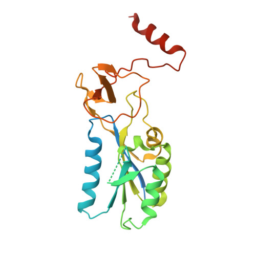
    Cytosolic thymidine kinase 1, TK1, is a well known cell-cycle-regulated enzyme of importance in nucleotide metabolism as well as an activator of antiviral and anticancer drugs such as 3'-azido-3'-deoxythymidine (AZT). We have now determined the structures of the TK1 family, the human and Ureaplasma urealyticum enzymes, in complex with the feedback inhibitor dTTP. The TK1s have a tetrameric structure in which each subunit contains an alpha/beta-domain that is similar to ATPase domains of members of the RecA structural family and a domain containing a structural zinc. The zinc ion connects beta-structures at the root of a beta-ribbon that forms a stem that widens to a lasso-type loop. The thymidine of dTTP is hydrogen-bonded to main-chain atoms predominantly coming from the lasso loop. This binding is in contrast to other deoxyribonucleoside kinases where specific interactions occur with side chains. The TK1 structure differs fundamentally from the structures of the other deoxyribonucleoside kinases, indicating a different evolutionary origin.


  • Organizational Affiliation

    Department of Molecular Biology, Swedish University of Agricultural Sciences, S-751 24 Uppsala, Sweden.


Macromolecules
Find similar proteins by:  (by identity cutoff)  |  3D Structure
Entity ID: 1
MoleculeChains Sequence LengthOrganismDetailsImage
THYMIDINE KINASE
A, B, C, D
243Ureaplasma urealyticumMutation(s): 1 
EC: 2.7.1.21
UniProt
Find proteins for Q9PPP5 (Ureaplasma parvum serovar 3 (strain ATCC 700970))
Explore Q9PPP5 
Go to UniProtKB:  Q9PPP5
Entity Groups  
Sequence Clusters30% Identity50% Identity70% Identity90% Identity95% Identity100% Identity
UniProt GroupQ9PPP5
Sequence Annotations
Expand
  • Reference Sequence
Small Molecules
Ligands 3 Unique
IDChains Name / Formula / InChI Key2D Diagram3D Interactions
TTP
Query on TTP

Download Ideal Coordinates CCD File 
F [auth A],
I [auth B],
K [auth C],
N [auth D]
THYMIDINE-5'-TRIPHOSPHATE
C10 H17 N2 O14 P3
NHVNXKFIZYSCEB-XLPZGREQSA-N
ZN
Query on ZN

Download Ideal Coordinates CCD File 
E [auth A],
G [auth B],
J [auth C],
L [auth D]
ZINC ION
Zn
PTFCDOFLOPIGGS-UHFFFAOYSA-N
MG
Query on MG

Download Ideal Coordinates CCD File 
H [auth B],
M [auth D]
MAGNESIUM ION
Mg
JLVVSXFLKOJNIY-UHFFFAOYSA-N
Experimental Data & Validation

Experimental Data

  • Method: X-RAY DIFFRACTION
  • Resolution: 2.50 Å
  • R-Value Free: 0.275 
  • R-Value Work: 0.220 
  • R-Value Observed: 0.223 
  • Space Group: P 1 21 1
Unit Cell:
Length ( Å )Angle ( ˚ )
a = 63.789α = 90
b = 110.753β = 109.86
c = 73.034γ = 90
Software Package:
Software NamePurpose
REFMACrefinement
DENZOdata reduction
SCALEPACKdata scaling
SHELXphasing

Structure Validation

View Full Validation Report


Ligand Structure Quality Assessment 


Entry History 

Deposition Data

Revision History  (Full details and data files)

  • Version 1.0: 2007-05-29
    Type: Initial release
  • Version 1.1: 2011-05-08
    Changes: Version format compliance
  • Version 1.2: 2011-07-13
    Changes: Version format compliance
  • Version 1.3: 2019-01-30
    Changes: Data collection, Experimental preparation, Other
  • Version 1.4: 2019-02-06
    Changes: Data collection, Experimental preparation