2UWA

Crystal structure of the Nasturtium seedling xyloglucanase isoform NXG1


Experimental Data Snapshot

  • Method: X-RAY DIFFRACTION
  • Resolution: 1.80 Å
  • R-Value Free: 0.200 
  • R-Value Work: 0.176 
  • R-Value Observed: 0.177 

wwPDB Validation   3D Report Full Report


This is version 1.3 of the entry. See complete history


Literature

Structural Evidence for the Evolution of Xyloglucanase Activity from Xyloglucan Endo-Transglycosylases: Biological Implications for Cell Wall Metabolism.

Baumann, M.J.Eklof, J.Michel, G.Kallasa, A.Teeri, T.T.Czjzek, M.Brumer, H.

(2007) Plant Cell 19: 1947

  • DOI: https://doi.org/10.1105/tpc.107.051391
  • Primary Citation of Related Structures:  
    2UWA, 2UWB, 2UWC

  • PubMed Abstract: 

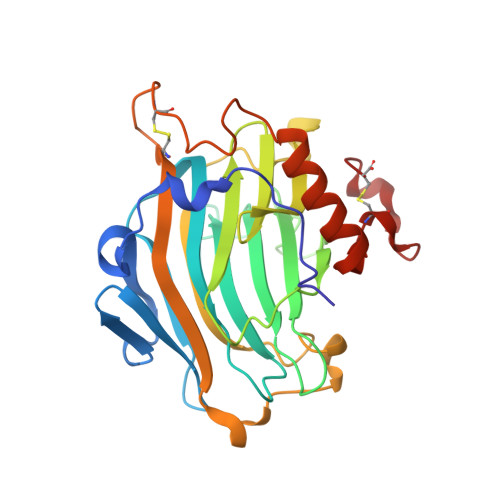
    High-resolution, three-dimensional structures of the archetypal glycoside hydrolase family 16 (GH16) endo-xyloglucanases Tm-NXG1 and Tm-NXG2 from nasturtium (Tropaeolum majus) have been solved by x-ray crystallography. Key structural features that modulate the relative rates of substrate hydrolysis to transglycosylation in the GH16 xyloglucan-active enzymes were identified by structure-function studies of the recombinantly expressed enzymes in comparison with data for the strict xyloglucan endo-transglycosylase Ptt-XET16-34 from hybrid aspen (Populus tremula x Populus tremuloides). Production of the loop deletion variant Tm-NXG1-DeltaYNIIG yielded an enzyme that was structurally similar to Ptt-XET16-34 and had a greatly increased transglycosylation:hydrolysis ratio. Comprehensive bioinformatic analyses of XTH gene products, together with detailed kinetic data, strongly suggest that xyloglucanase activity has evolved as a gain of function in an ancestral GH16 XET to meet specific biological requirements during seed germination, fruit ripening, and rapid wall expansion.


  • Organizational Affiliation

    School of Biotechnology, Royal Institute of Technology, AlbaNova University Center, Stockholm, Sweden.


Macromolecules
Find similar proteins by:  (by identity cutoff)  |  3D Structure
Entity ID: 1
MoleculeChains Sequence LengthOrganismDetailsImage
CELLULASE
A, B, C
274Tropaeolum majusMutation(s): 0 
EC: 3.2.1.151 (PDB Primary Data), 3.2.1.4 (PDB Primary Data)
UniProt
Find proteins for Q07524 (Tropaeolum majus)
Explore Q07524 
Go to UniProtKB:  Q07524
Entity Groups  
Sequence Clusters30% Identity50% Identity70% Identity90% Identity95% Identity100% Identity
UniProt GroupQ07524
Sequence Annotations
Expand
  • Reference Sequence
Experimental Data & Validation

Experimental Data

  • Method: X-RAY DIFFRACTION
  • Resolution: 1.80 Å
  • R-Value Free: 0.200 
  • R-Value Work: 0.176 
  • R-Value Observed: 0.177 
  • Space Group: P 31
Unit Cell:
Length ( Å )Angle ( ˚ )
a = 116.109α = 90
b = 116.109β = 90
c = 63.071γ = 120
Software Package:
Software NamePurpose
REFMACrefinement
MOSFLMdata reduction
SCALAdata scaling
AMoREphasing

Structure Validation

View Full Validation Report


Entry History 

Deposition Data

Revision History  (Full details and data files)

  • Version 1.0: 2007-06-26
    Type: Initial release
  • Version 1.1: 2011-05-08
    Changes: Version format compliance
  • Version 1.2: 2011-07-13
    Changes: Version format compliance
  • Version 1.3: 2023-12-13
    Changes: Data collection, Database references, Derived calculations, Other, Refinement description