2RMP

RMP-pepstatin A complex


Experimental Data Snapshot

  • Method: X-RAY DIFFRACTION
  • Resolution: 2.70 Å
  • R-Value Free: 0.280 
  • R-Value Work: 0.193 
  • R-Value Observed: 0.193 

wwPDB Validation   3D Report Full Report


This is version 2.1 of the entry. See complete history


Literature

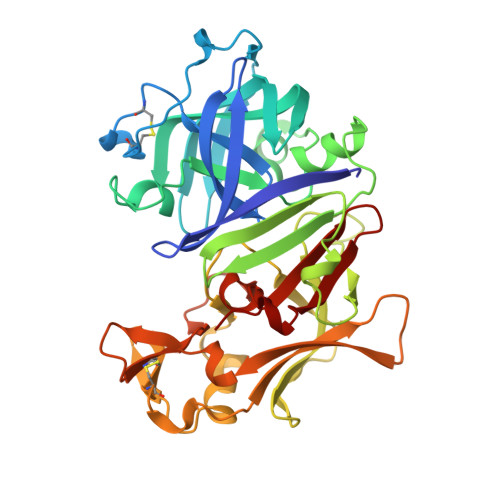
Structure of the Rhizomucor miehei aspartic proteinase complexed with the inhibitor pepstatin A at 2.7 A resolution.

Yang, J.Quail, J.W.

(1999) Acta Crystallogr D Biol Crystallogr 55: 625-630

  • DOI: https://doi.org/10.1107/s0907444998013961
  • Primary Citation of Related Structures:  
    2RMP

  • PubMed Abstract: 

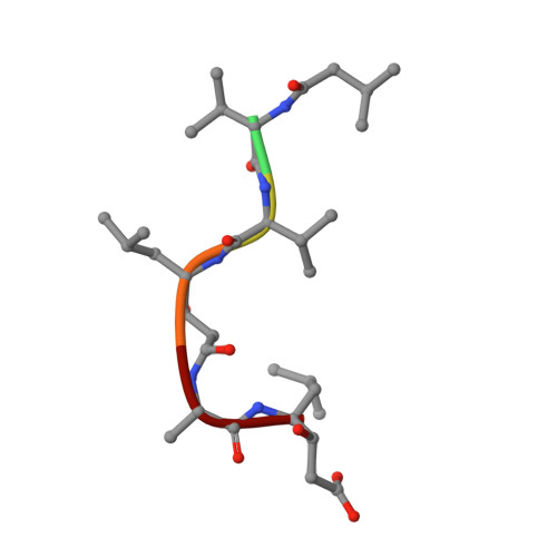
    Crystals of Rhizomucor miehei aspartic proteinase (RMP) complexed with pepstatin A grew in the orthorhombic space group P212121 and were isomorphous to native RMP crystals. The unit-cell dimensions are a = 41.52, b = 50.82, c = 172.71 A. There is one RMP-pepstatin A complex per asymmetric unit. The structure of the RMP-pepstatin A complex has been refined to a crystallographic R value of 19.3% and an Rfree value of 28.0% at 2.7 A resolution. A pepstatin A molecule fits into the large substrate-binding cleft between the two domains of RMP in an extended conformation up to the alanine residue at the P2' position. The dipeptide analogue statine residue at the P3'-P4' position forms an inverse gamma-turn (P3'-P1') with the statine residue at the P1-P1' position and its leucyl side chain binds back into the S1' subsite. The inhibitor interacts with the residues of the substrate-binding pocket by both hydrogen bonds and hydrophobic interactions. The hydroxyl group of the statine residue at the P1-P1' position forms hydrogen bonds with both catalytic aspartate residues (Asp38 and Asp237). This conformation mimics the expected transition state of the enzyme-substrate interaction. The binding of the inhibitor to the enzyme does not produce large distortions of the active site. No domain movement was observed compared with the native enzyme structure. However, the surface-flap region (residues 82-88) undergoes a conformational change, moving toward the inhibitor and becoming rigid owing to the formation of hydrogen bonds with the inhibitor. B-factor calculations of the two domains suggest that the C-terminal domain becomes more rigid in the complex than in the native structure.


  • Organizational Affiliation

    Department of Chemistry, University of Saskatchewan, 110 Science Place, Saskatoon, Saskatchewan S7N 5C9, Canada.


Macromolecules
Find similar proteins by:  (by identity cutoff)  |  3D Structure
Entity ID: 1
MoleculeChains Sequence LengthOrganismDetailsImage
MUCOROPEPSIN361Rhizomucor mieheiMutation(s): 0 
EC: 3.4.23.23
UniProt
Find proteins for P00799 (Rhizomucor miehei)
Explore P00799 
Go to UniProtKB:  P00799
Entity Groups  
Sequence Clusters30% Identity50% Identity70% Identity90% Identity95% Identity100% Identity
UniProt GroupP00799
Sequence Annotations
Expand
  • Reference Sequence

Find similar proteins by:  Sequence   |   3D Structure  

Entity ID: 2
MoleculeChains Sequence LengthOrganismDetailsImage
PEPSTATIN6StreptomycesMutation(s): 0 
Sequence Annotations
Expand
  • Reference Sequence
Oligosaccharides

Help

Entity ID: 3
MoleculeChains Length2D Diagram Glycosylation3D Interactions
beta-D-mannopyranose-(1-4)-2-acetamido-2-deoxy-beta-D-glucopyranose
C
2N-Glycosylation
Glycosylation Resources
GlyTouCan:  G90333CG
GlyCosmos:  G90333CG
GlyGen:  G90333CG
Small Molecules
Ligands 1 Unique
IDChains Name / Formula / InChI Key2D Diagram3D Interactions
NAG
Query on NAG

Download Ideal Coordinates CCD File 
D [auth A]2-acetamido-2-deoxy-beta-D-glucopyranose
C8 H15 N O6
OVRNDRQMDRJTHS-FMDGEEDCSA-N
Biologically Interesting Molecules (External Reference) 1 Unique
Entity ID: 2
IDChains NameType/Class2D Diagram3D Interactions
PRD_000557
Query on PRD_000557
B
PepstatinOligopeptide / Enzyme inhibitor
Experimental Data & Validation

Experimental Data

  • Method: X-RAY DIFFRACTION
  • Resolution: 2.70 Å
  • R-Value Free: 0.280 
  • R-Value Work: 0.193 
  • R-Value Observed: 0.193 
  • Space Group: P 21 21 21
Unit Cell:
Length ( Å )Angle ( ˚ )
a = 41.52α = 90
b = 50.82β = 90
c = 172.71γ = 90
Software Package:
Software NamePurpose
DENZOdata reduction
SCALEPACKdata scaling
X-PLORmodel building
X-PLORrefinement
X-PLORphasing

Structure Validation

View Full Validation Report


Entry History 

Deposition Data

Revision History  (Full details and data files)

  • Version 1.0: 1997-09-17
    Type: Initial release
  • Version 1.1: 2008-03-25
    Changes: Version format compliance
  • Version 1.2: 2011-07-13
    Changes: Atomic model, Database references, Derived calculations, Non-polymer description, Structure summary, Version format compliance
  • Version 1.3: 2013-02-27
    Changes: Other
  • Version 1.4: 2018-04-18
    Changes: Data collection
  • Version 2.0: 2020-07-29
    Type: Remediation
    Reason: Carbohydrate remediation
    Changes: Atomic model, Data collection, Derived calculations, Structure summary
  • Version 2.1: 2023-08-09
    Changes: Database references, Refinement description, Structure summary