2QMI

Structure of the octameric penicillin-binding protein homologue from Pyrococcus abyssi


Experimental Data Snapshot

  • Method: X-RAY DIFFRACTION
  • Resolution: 2.20 Å
  • R-Value Free: 0.241 
  • R-Value Work: 0.196 
  • R-Value Observed: 0.196 

wwPDB Validation   3D Report Full Report


Ligand Structure Quality Assessment 


This is version 1.2 of the entry. See complete history


Literature

Structure of the archaeal pab87 peptidase reveals a novel self-compartmentalizing protease family

Delfosse, V.Girard, E.Birck, C.Delmarcelle, M.Delarue, M.Poch, O.Schultz, P.Mayer, C.

(2009) PLoS One 4: e4712-e4712

  • DOI: https://doi.org/10.1371/journal.pone.0004712
  • Primary Citation of Related Structures:  
    2QMI

  • PubMed Abstract: 

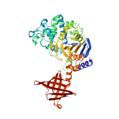
    Self-compartmentalizing proteases orchestrate protein turnover through an original architecture characterized by a central catalytic chamber. Here we report the first structure of an archaeal member of a new self-compartmentalizing protease family forming a cubic-shaped octamer with D(4) symmetry and referred to as CubicO. We solved the structure of the Pyrococcus abyssi Pab87 protein at 2.2 A resolution using the anomalous signal of the high-phasing-power lanthanide derivative Lu-HPDO3A. A 20 A wide channel runs through this supramolecular assembly of 0.4 MDa, giving access to a 60 A wide central chamber holding the eight active sites. Surprisingly, activity assays revealed that Pab87 degrades specifically d-amino acid containing peptides, which have never been observed in archaea. Genomic context of the Pab87 gene showed that it is surrounded by genes involved in the amino acid/peptide transport or metabolism. We propose that CubicO proteases are involved in the processing of d-peptides from environmental origins.


  • Organizational Affiliation

    Centre de Recherche des Cordeliers, LRMA, INSERM UMR-S 872, Université Pierre et Marie Curie, Paris, France.


Macromolecules
Find similar proteins by:  (by identity cutoff)  |  3D Structure
Entity ID: 1
MoleculeChains Sequence LengthOrganismDetailsImage
Pbp related beta-lactamase
A, B, C, D, E
A, B, C, D, E, F, G, H
447Pyrococcus abyssiMutation(s): 0 
Gene Names: pbp
EC: 3.5.2.6
UniProt
Find proteins for Q9V2D6 (Pyrococcus abyssi (strain GE5 / Orsay))
Explore Q9V2D6 
Go to UniProtKB:  Q9V2D6
Entity Groups  
Sequence Clusters30% Identity50% Identity70% Identity90% Identity95% Identity100% Identity
UniProt GroupQ9V2D6
Sequence Annotations
Expand
  • Reference Sequence
Small Molecules
Ligands 2 Unique
IDChains Name / Formula / InChI Key2D Diagram3D Interactions
DO3
Query on DO3

Download Ideal Coordinates CCD File 
AA [auth G],
CA [auth H],
S [auth E],
W [auth F]
10-((2R)-2-HYDROXYPROPYL)-1,4,7,10-TETRAAZACYCLODODECANE 1,4,7-TRIACETIC ACID
C17 H32 N4 O7
IQUHNCOJRJBMSU-CQSZACIVSA-N
LU
Query on LU

Download Ideal Coordinates CCD File 
BA [auth H]
I [auth A]
J [auth A]
K [auth B]
L [auth B]
BA [auth H],
I [auth A],
J [auth A],
K [auth B],
L [auth B],
M [auth C],
N [auth C],
O [auth D],
P [auth D],
Q [auth E],
R [auth E],
T [auth F],
U [auth F],
V [auth F],
X [auth G],
Y [auth G],
Z [auth G]
LUTETIUM (III) ION
Lu
PSDMOPINLDTFSZ-UHFFFAOYSA-N
Experimental Data & Validation

Experimental Data

  • Method: X-RAY DIFFRACTION
  • Resolution: 2.20 Å
  • R-Value Free: 0.241 
  • R-Value Work: 0.196 
  • R-Value Observed: 0.196 
  • Space Group: P 1
Unit Cell:
Length ( Å )Angle ( ˚ )
a = 100.94α = 72.17
b = 105.63β = 66.51
c = 112.92γ = 81.39
Software Package:
Software NamePurpose
CNSrefinement
PDB_EXTRACTdata extraction
ADSCdata collection
XDSdata reduction
SCALAdata scaling
SHARPphasing

Structure Validation

View Full Validation Report


Ligand Structure Quality Assessment 


Entry History 

Deposition Data

Revision History  (Full details and data files)

  • Version 1.0: 2008-07-22
    Type: Initial release
  • Version 1.1: 2011-07-13
    Changes: Version format compliance
  • Version 1.2: 2024-02-21
    Changes: Data collection, Database references, Derived calculations