2QLL

Human liver glycogen phosphorylase- GL complex


Experimental Data Snapshot

  • Method: X-RAY DIFFRACTION
  • Resolution: 2.56 Å
  • R-Value Free: 0.276 
  • R-Value Work: 0.203 
  • R-Value Observed: 0.206 

wwPDB Validation   3D Report Full Report


Ligand Structure Quality Assessment 


This is version 1.3 of the entry. See complete history


Literature

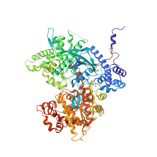
Molecular recognition of the protein phosphatase 1 glycogen targeting subunit by glycogen phosphorylase.

Pautsch, A.Stadler, N.Wissdorf, O.Langkopf, E.Moreth, W.Streicher, R.

(2008) J Biol Chem 283: 8913-8918

  • DOI: https://doi.org/10.1074/jbc.M706612200
  • Primary Citation of Related Structures:  
    2QLL

  • PubMed Abstract: 

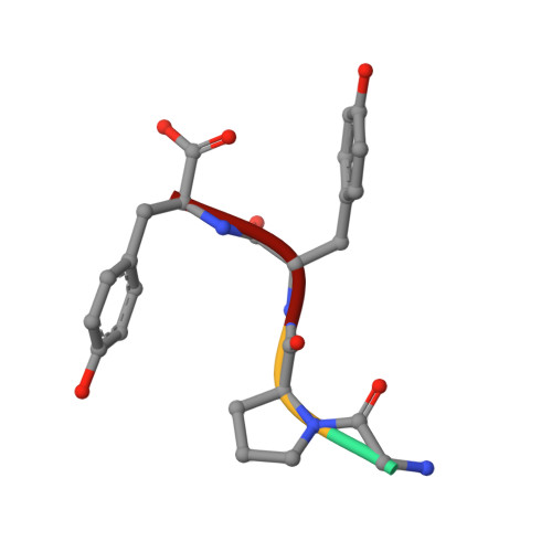
    Disrupting the interaction between glycogen phosphorylase and the glycogen targeting subunit (G(L)) of protein phosphatase 1 is emerging as a novel target for the treatment of type 2 diabetes. To elucidate the molecular basis of binding, we have determined the crystal structure of liver phosphorylase bound to a G(L)-derived peptide. The structure reveals the C terminus of G(L) binding in a hydrophobically collapsed conformation to the allosteric regulator-binding site at the phosphorylase dimer interface. G(L) mimics interactions that are otherwise employed by the activator AMP. Functional studies show that G(L) binds tighter than AMP and confirm that the C-terminal Tyr-Tyr motif is the major determinant for G(L) binding potency. Our study validates the G(L)-phosphorylase interface as a novel target for small molecule interaction.


  • Organizational Affiliation

    Department of Lead Discovery, Boehringer Ingelheim GmbH & Co. KG, Birkendorferstrasse 65, Biberach, Germany. alexander.pautsch@boehringer-ingelheim.com


Macromolecules
Find similar proteins by:  (by identity cutoff)  |  3D Structure
Entity ID: 1
MoleculeChains Sequence LengthOrganismDetailsImage
Glycogen phosphorylase, liver form847Homo sapiensMutation(s): 1 
Gene Names: PYGL
EC: 2.4.1.1
UniProt & NIH Common Fund Data Resources
Find proteins for P06737 (Homo sapiens)
Explore P06737 
Go to UniProtKB:  P06737
PHAROS:  P06737
GTEx:  ENSG00000100504 
Entity Groups  
Sequence Clusters30% Identity50% Identity70% Identity90% Identity95% Identity100% Identity
UniProt GroupP06737
Sequence Annotations
Expand
  • Reference Sequence

Find similar proteins by:  Sequence   |   3D Structure  

Entity ID: 2
MoleculeChains Sequence LengthOrganismDetailsImage
protein targeting to glycogen - GL4N/AMutation(s): 0 
Sequence Annotations
Expand
  • Reference Sequence
Small Molecules
Ligands 1 Unique
IDChains Name / Formula / InChI Key2D Diagram3D Interactions
PLP
Query on PLP

Download Ideal Coordinates CCD File 
C [auth A]PYRIDOXAL-5'-PHOSPHATE
C8 H10 N O6 P
NGVDGCNFYWLIFO-UHFFFAOYSA-N
Modified Residues  1 Unique
IDChains TypeFormula2D DiagramParent
SEP
Query on SEP
A
L-PEPTIDE LINKINGC3 H8 N O6 PSER
Experimental Data & Validation

Experimental Data

  • Method: X-RAY DIFFRACTION
  • Resolution: 2.56 Å
  • R-Value Free: 0.276 
  • R-Value Work: 0.203 
  • R-Value Observed: 0.206 
  • Space Group: P 31 2 1
Unit Cell:
Length ( Å )Angle ( ˚ )
a = 123.986α = 90
b = 123.986β = 90
c = 125.695γ = 120
Software Package:
Software NamePurpose
XSCALEdata scaling
REFMACrefinement
PDB_EXTRACTdata extraction
XDSdata reduction
BUSTER-TNTrefinement

Structure Validation

View Full Validation Report


Ligand Structure Quality Assessment 


Entry History 

Deposition Data

Revision History  (Full details and data files)

  • Version 1.0: 2008-02-19
    Type: Initial release
  • Version 1.1: 2011-07-13
    Changes: Non-polymer description, Version format compliance
  • Version 1.2: 2012-12-12
    Changes: Other
  • Version 1.3: 2013-03-13
    Changes: Other