2QJU

Crystal Structure of an NSS Homolog with Bound Antidepressant


Experimental Data Snapshot

  • Method: X-RAY DIFFRACTION
  • Resolution: 2.90 Å
  • R-Value Free: 0.220 
  • R-Value Work: 0.199 
  • R-Value Observed: 0.199 

wwPDB Validation   3D Report Full Report


Ligand Structure Quality Assessment 


This is version 1.3 of the entry. See complete history


Literature

LeuT-desipramine structure reveals how antidepressants block neurotransmitter reuptake.

Zhou, Z.Zhen, J.Karpowich, N.K.Goetz, R.M.Law, C.J.Reith, M.E.Wang, D.N.

(2007) Science 317: 1390-1393

  • DOI: https://doi.org/10.1126/science.1147614
  • Primary Citation of Related Structures:  
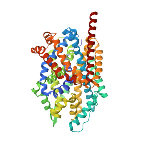
    2QJU

  • PubMed Abstract: 

    Tricyclic antidepressants exert their pharmacological effect-inhibiting the reuptake of serotonin, norepinephrine, and dopamine-by directly blocking neurotransmitter transporters (SERT, NET, and DAT, respectively) in the presynaptic membrane. The drug-binding site and the mechanism of this inhibition are poorly understood. We determined the crystal structure at 2.9 angstroms of the bacterial leucine transporter (LeuT), a homolog of SERT, NET, and DAT, in complex with leucine and the antidepressant desipramine. Desipramine binds at the inner end of the extracellular cavity of the transporter and is held in place by a hairpin loop and by a salt bridge. This binding site is separated from the leucine-binding site by the extracellular gate of the transporter. By directly locking the gate, desipramine prevents conformational changes and blocks substrate transport. Mutagenesis experiments on human SERT and DAT indicate that both the desipramine-binding site and its inhibition mechanism are probably conserved in the human neurotransmitter transporters.


  • Organizational Affiliation

    Kimmel Center for Biology and Medicine at the Skirball Institute of Biomolecular Medicine and Department of Cell Biology, New York University School of Medicine, 540 First Avenue, New York, NY 10016, USA.


Macromolecules
Find similar proteins by:  (by identity cutoff)  |  3D Structure
Entity ID: 1
MoleculeChains Sequence LengthOrganismDetailsImage
Transporter511Aquifex aeolicus VF5Mutation(s): 0 
Gene Names: snf
Membrane Entity: Yes 
UniProt
Find proteins for O67854 (Aquifex aeolicus (strain VF5))
Explore O67854 
Go to UniProtKB:  O67854
Entity Groups  
Sequence Clusters30% Identity50% Identity70% Identity90% Identity95% Identity100% Identity
UniProt GroupO67854
Sequence Annotations
Expand
  • Reference Sequence
Small Molecules
Ligands 5 Unique
IDChains Name / Formula / InChI Key2D Diagram3D Interactions
BOG
Query on BOG

Download Ideal Coordinates CCD File 
B [auth A],
C [auth A],
D [auth A],
E [auth A]
octyl beta-D-glucopyranoside
C14 H28 O6
HEGSGKPQLMEBJL-RKQHYHRCSA-N
DSM
Query on DSM

Download Ideal Coordinates CCD File 
J [auth A]3-(10,11-DIHYDRO-5H-DIBENZO[B,F]AZEPIN-5-YL)-N-METHYLPROPAN-1-AMINE
C18 H22 N2
HCYAFALTSJYZDH-UHFFFAOYSA-N
LEU
Query on LEU

Download Ideal Coordinates CCD File 
I [auth A]LEUCINE
C6 H13 N O2
ROHFNLRQFUQHCH-YFKPBYRVSA-N
CL
Query on CL

Download Ideal Coordinates CCD File 
H [auth A]CHLORIDE ION
Cl
VEXZGXHMUGYJMC-UHFFFAOYSA-M
NA
Query on NA

Download Ideal Coordinates CCD File 
F [auth A],
G [auth A]
SODIUM ION
Na
FKNQFGJONOIPTF-UHFFFAOYSA-N
Binding Affinity Annotations 
IDSourceBinding Affinity
DSM PDBBind:  2QJU IC50: 8.00e+4 (nM) from 1 assay(s)
Experimental Data & Validation

Experimental Data

  • Method: X-RAY DIFFRACTION
  • Resolution: 2.90 Å
  • R-Value Free: 0.220 
  • R-Value Work: 0.199 
  • R-Value Observed: 0.199 
  • Space Group: C 1 2 1
Unit Cell:
Length ( Å )Angle ( ˚ )
a = 88.414α = 90
b = 86.763β = 95.72
c = 81.187γ = 90
Software Package:
Software NamePurpose
CNSrefinement
HKL-2000data collection
HKL-2000data reduction
HKL-2000data scaling
CNSphasing

Structure Validation

View Full Validation Report


Ligand Structure Quality Assessment 


Entry History 

Deposition Data

Revision History  (Full details and data files)

  • Version 1.0: 2007-08-21
    Type: Initial release
  • Version 1.1: 2011-07-13
    Changes: Source and taxonomy, Version format compliance
  • Version 1.2: 2020-07-29
    Type: Remediation
    Reason: Carbohydrate remediation
    Changes: Data collection, Database references, Derived calculations, Structure summary
  • Version 1.3: 2023-08-30
    Changes: Data collection, Database references, Refinement description, Structure summary