2Q9B

Structure of FTSY:GMPPNP Complex


Experimental Data Snapshot

  • Method: X-RAY DIFFRACTION
  • Resolution: 2.30 Å
  • R-Value Free: 0.269 
  • R-Value Work: 0.208 
  • R-Value Observed: 0.208 

wwPDB Validation   3D Report Full Report


Ligand Structure Quality Assessment 


This is version 1.3 of the entry. See complete history


Literature

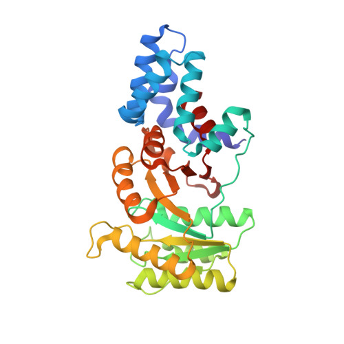
X-ray Structures of the Signal Recognition Particle Receptor Reveal Targeting Cycle Intermediates.

Reyes, C.L.Rutenber, E.Walter, P.Stroud, R.M.

(2007) PLoS One 2: e607-e607

  • DOI: https://doi.org/10.1371/journal.pone.0000607
  • Primary Citation of Related Structures:  
    2Q9A, 2Q9B, 2Q9C

  • PubMed Abstract: 

    The signal recognition particle (SRP) and its conjugate receptor (SR) mediate cotranslational targeting of a subclass of proteins destined for secretion to the endoplasmic reticulum membrane in eukaryotes or to the plasma membrane in prokaryotes. Conserved active site residues in the GTPase domains of both SRP and SR mediate discrete conformational changes during formation and dissociation of the SRP.SR complex. Here, we describe structures of the prokaryotic SR, FtsY, as an apo protein and in two different complexes with a non-hydrolysable GTP analog (GMPPNP). These structures reveal intermediate conformations of FtsY containing GMPPNP and explain how the conserved active site residues position the nucleotide into a non-catalytic conformation. The basis for the lower specificity of binding of nucleotide in FtsY prior to heterodimerization with the SRP conjugate Ffh is also shown. We propose that these structural changes represent discrete conformational states assumed by FtsY during targeting complex formation and dissociation.


  • Organizational Affiliation

    Graduate Group in Biophysics, Department of Biochemistry and Biophysics, University of California at San Francisco, San Francisco, California, United States of America.


Macromolecules
Find similar proteins by:  (by identity cutoff)  |  3D Structure
Entity ID: 1
MoleculeChains Sequence LengthOrganismDetailsImage
Cell division protein ftsY
A, B
304Thermus aquaticusMutation(s): 0 
Gene Names: ftsY
UniProt
Find proteins for P83749 (Thermus aquaticus)
Explore P83749 
Go to UniProtKB:  P83749
Entity Groups  
Sequence Clusters30% Identity50% Identity70% Identity90% Identity95% Identity100% Identity
UniProt GroupP83749
Sequence Annotations
Expand
  • Reference Sequence
Small Molecules
Ligands 3 Unique
IDChains Name / Formula / InChI Key2D Diagram3D Interactions
GNP
Query on GNP

Download Ideal Coordinates CCD File 
L [auth A]PHOSPHOAMINOPHOSPHONIC ACID-GUANYLATE ESTER
C10 H17 N6 O13 P3
UQABYHGXWYXDTK-UUOKFMHZSA-N
SO4
Query on SO4

Download Ideal Coordinates CCD File 
C [auth A]
D [auth A]
E [auth A]
F [auth A]
G [auth A]
C [auth A],
D [auth A],
E [auth A],
F [auth A],
G [auth A],
H [auth A],
M [auth B],
N [auth B]
SULFATE ION
O4 S
QAOWNCQODCNURD-UHFFFAOYSA-L
EDO
Query on EDO

Download Ideal Coordinates CCD File 
I [auth A]
J [auth A]
K [auth A]
O [auth B]
P [auth B]
I [auth A],
J [auth A],
K [auth A],
O [auth B],
P [auth B],
Q [auth B],
R [auth B],
S [auth B],
T [auth B]
1,2-ETHANEDIOL
C2 H6 O2
LYCAIKOWRPUZTN-UHFFFAOYSA-N
Experimental Data & Validation

Experimental Data

  • Method: X-RAY DIFFRACTION
  • Resolution: 2.30 Å
  • R-Value Free: 0.269 
  • R-Value Work: 0.208 
  • R-Value Observed: 0.208 
  • Space Group: P 21 21 21
Unit Cell:
Length ( Å )Angle ( ˚ )
a = 63.843α = 90
b = 97.298β = 90
c = 99.284γ = 90
Software Package:
Software NamePurpose
CNSrefinement

Structure Validation

View Full Validation Report


Ligand Structure Quality Assessment 


Entry History 

Deposition Data

Revision History  (Full details and data files)

  • Version 1.0: 2007-07-03
    Type: Initial release
  • Version 1.1: 2008-05-01
    Changes: Version format compliance
  • Version 1.2: 2011-07-13
    Changes: Derived calculations, Version format compliance
  • Version 1.3: 2024-02-21
    Changes: Data collection, Database references, Derived calculations