Structural insight into substrate binding and catalysis of a novel 2-keto-3-deoxy-D-arabinonate dehydratase illustrates common mechanistic features of the FAH superfamily.
Brouns, S.J., Barends, T.R., Worm, P., Akerboom, J., Turnbull, A.P., Salmon, L., van der Oost, J.(2008) J Mol Biol 379: 357-371
- PubMed: 18448118
- DOI: https://doi.org/10.1016/j.jmb.2008.03.064
- PubMed Abstract:
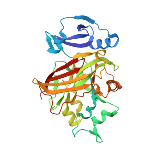
The archaeon Sulfolobus solfataricus converts d-arabinose to 2-oxoglutarate by an enzyme set consisting of two dehydrogenases and two dehydratases. The third step of the pathway is catalyzed by a novel 2-keto-3-deoxy-D-arabinonate dehydratase (KdaD). In this study, the crystal structure of the enzyme has been solved to 2.1 A resolution. The enzyme forms an oval-shaped ring of four subunits, each consisting of an N-terminal domain with a four-stranded beta-sheet flanked by two alpha-helices, and a C-terminal catalytic domain with a fumarylacetoacetate hydrolase (FAH) fold. Crystal structures of complexes of the enzyme with magnesium or calcium ions and either a substrate analog 2-oxobutyrate, or the aldehyde enzyme product 2,5-dioxopentanoate revealed that the divalent metal ion in the active site is coordinated octahedrally by three conserved carboxylate residues, a water molecule, and both the carboxylate and the oxo groups of the substrate molecule. An enzymatic mechanism for base-catalyzed dehydration is proposed on the basis of the binding mode of the substrate to the metal ion, which suggests that the enzyme enhances the acidity of the protons alpha to the carbonyl group, facilitating their abstraction by glutamate 114. A comprehensive structural comparison of members of the FAH superfamily is presented and their evolution is discussed, providing a basis for functional investigations of this largely unexplored protein superfamily.
Organizational Affiliation:
Laboratory of Microbiology, Department of Agrotechnology and Food Sciences, Wageningen University, Dreienplein 10, 6703 HB Wageningen, Netherlands. stan.brouns@wur.nl