2Q12

Crystal Structure of BAR domain of APPL1


Experimental Data Snapshot

  • Method: X-RAY DIFFRACTION
  • Resolution: 1.79 Å
  • R-Value Free: 0.255 
  • R-Value Work: 0.212 
  • R-Value Observed: 0.214 

wwPDB Validation   3D Report Full Report


This is version 1.1 of the entry. See complete history


Literature

Structure of the APPL1 BAR-PH domain and characterization of its interaction with Rab5.

Zhu, G.Chen, J.Liu, J.Brunzelle, J.S.Huang, B.Wakeham, N.Terzyan, S.Li, X.Rao, Z.Li, G.Zhang, X.C.

(2007) EMBO J 26: 3484-3493

  • DOI: https://doi.org/10.1038/sj.emboj.7601771
  • Primary Citation of Related Structures:  
    2Q12, 2Q13

  • PubMed Abstract: 

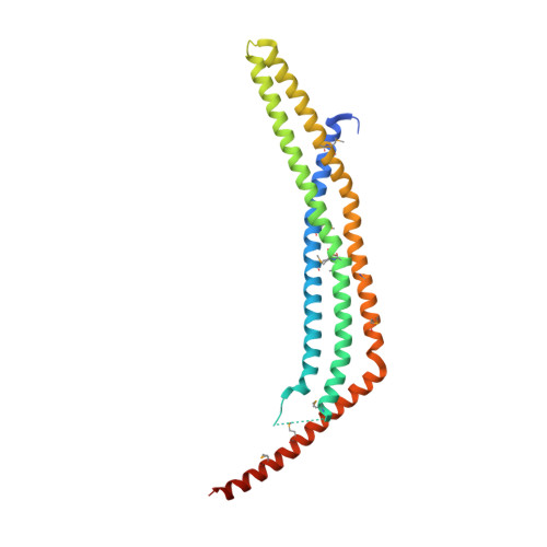
    APPL1 is an effector of the small GTPase Rab5. Together, they mediate a signal transduction pathway initiated by ligand binding to cell surface receptors. Interaction with Rab5 is confined to the amino (N)-terminal region of APPL1. We report the crystal structures of human APPL1 N-terminal BAR-PH domain motif. The BAR and PH domains, together with a novel linker helix, form an integrated, crescent-shaped, symmetrical dimer. This BAR-PH interaction is likely conserved in the class of BAR-PH containing proteins. Biochemical analyses indicate two independent Rab-binding sites located at the opposite ends of the dimer, where the PH domain directly interacts with Rab5 and Rab21. Besides structurally supporting the PH domain, the BAR domain also contributes to Rab binding through a small surface region in the vicinity of the PH domain. In stark contrast to the helix-dominated, Rab-binding domains previously reported, APPL1 PH domain employs beta-strands to interact with Rab5. On the Rab5 side, both switch regions are involved in the interaction. Thus we identified a new binding mode between PH domains and small GTPases.


  • Organizational Affiliation

    Crystallography Research Program, Oklahoma Medical Research Foundation, Oklahoma City, OK 73104, USA.


Macromolecules
Find similar proteins by:  (by identity cutoff)  |  3D Structure
Entity ID: 1
MoleculeChains Sequence LengthOrganismDetailsImage
DCC-interacting protein 13 alpha265Homo sapiensMutation(s): 0 
Gene Names: APPL1APPLDIP13AKIAA1428
UniProt & NIH Common Fund Data Resources
Find proteins for Q9UKG1 (Homo sapiens)
Explore Q9UKG1 
Go to UniProtKB:  Q9UKG1
PHAROS:  Q9UKG1
GTEx:  ENSG00000157500 
Entity Groups  
Sequence Clusters30% Identity50% Identity70% Identity90% Identity95% Identity100% Identity
UniProt GroupQ9UKG1
Sequence Annotations
Expand
  • Reference Sequence
Small Molecules
Modified Residues  1 Unique
IDChains TypeFormula2D DiagramParent
MSE
Query on MSE
A
L-PEPTIDE LINKINGC5 H11 N O2 SeMET
Experimental Data & Validation

Experimental Data

  • Method: X-RAY DIFFRACTION
  • Resolution: 1.79 Å
  • R-Value Free: 0.255 
  • R-Value Work: 0.212 
  • R-Value Observed: 0.214 
  • Space Group: P 21 21 2
Unit Cell:
Length ( Å )Angle ( ˚ )
a = 52.985α = 90
b = 129.199β = 90
c = 36.854γ = 90
Software Package:
Software NamePurpose
REFMACrefinement
HKL-2000data collection
HKL-2000data reduction
HKL-2000data scaling
SHARPphasing

Structure Validation

View Full Validation Report


Entry History 

Deposition Data

Revision History  (Full details and data files)

  • Version 1.0: 2007-08-14
    Type: Initial release
  • Version 1.1: 2011-07-13
    Changes: Derived calculations, Version format compliance