2PVV

Structure of human glutamate carboxypeptidase II (GCPII) in complex with L-serine-O-sulfate


Experimental Data Snapshot

  • Method: X-RAY DIFFRACTION
  • Resolution: 2.11 Å
  • R-Value Free: 0.212 
  • R-Value Work: 0.174 
  • R-Value Observed: 0.176 

wwPDB Validation   3D Report Full Report


Ligand Structure Quality Assessment 


This is version 2.1 of the entry. See complete history


Literature

Structural insight into the pharmacophore pocket of human glutamate carboxypeptidase II.

Barinka, C.Rovenska, M.Mlcochova, P.Hlouchova, K.Plechanovova, A.Majer, P.Tsukamoto, T.Slusher, B.S.Konvalinka, J.Lubkowski, J.

(2007) J Med Chem 50: 3267-3273

  • DOI: https://doi.org/10.1021/jm070133w
  • Primary Citation of Related Structures:  
    2OR4, 2PVV, 2PVW

  • PubMed Abstract: 

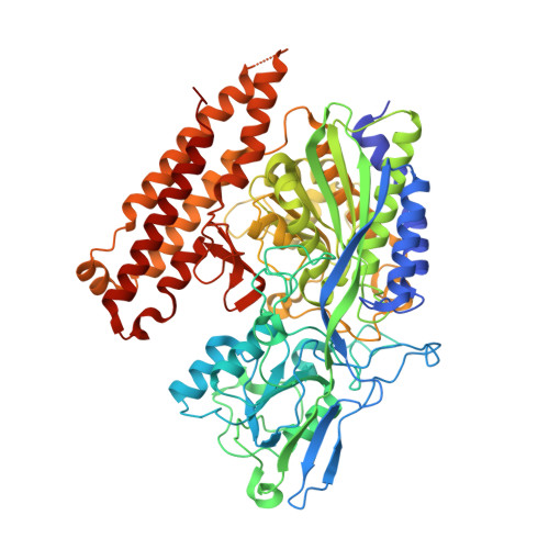
    Inhibition of glutamate carboxypeptidase II (GCPII) has been shown to be neuroprotective in multiple preclinical models in which dysregulated glutamatergic transmission is implicated. Herein, we report crystal structures of the human GCPII complexed with three glutamate mimetics/derivatives, 2-(phosphonomethyl)pentanedioic acid (2-PMPA), quisqualic acid (QA), and L-serine O-sulfate (L-SOS), at 1.72, 1.62, and 2.10 A resolution, respectively. Despite the structural differences between the distal parts of the inhibitors, all three compounds share similar binding modes in the pharmacophore (i.e., S1') pocket of GCPII, where they are stabilized by a combination of polar and van der Waals interactions. The structural diversity of the distal parts of the inhibitors leads to rearrangements of the S1' site that are necessary for efficient interactions between the enzyme and an inhibitor. The set of structures presented here, in conjunction with the available biochemical data, illustrates a flexibility of the GCPII pharmacophore pocket and highlights the structural features required for potent GCPII inhibition. These findings could facilitate the rational structure-based drug design of new GCPII inhibitors in the future.


  • Organizational Affiliation

    Center for Cancer Research, National Cancer Institute at Frederick, Frederick, Maryland 21702, USA.


Macromolecules
Find similar proteins by:  (by identity cutoff)  |  3D Structure
Entity ID: 1
MoleculeChains Sequence LengthOrganismDetailsImage
Glutamate carboxypeptidase 2709Homo sapiensMutation(s): 0 
Gene Names: FOLH1FOLHNAALAD1PSMPSMA
EC: 3.4.17.21
UniProt & NIH Common Fund Data Resources
Find proteins for Q04609 (Homo sapiens)
Explore Q04609 
Go to UniProtKB:  Q04609
PHAROS:  Q04609
GTEx:  ENSG00000086205 
Entity Groups  
Sequence Clusters30% Identity50% Identity70% Identity90% Identity95% Identity100% Identity
UniProt GroupQ04609
Sequence Annotations
Expand
  • Reference Sequence
Oligosaccharides

Help

Entity ID: 2
MoleculeChains Length2D Diagram Glycosylation3D Interactions
2-acetamido-2-deoxy-beta-D-glucopyranose-(1-4)-2-acetamido-2-deoxy-beta-D-glucopyranose
B, C, D
2N-Glycosylation
Glycosylation Resources
GlyTouCan:  G42666HT
GlyCosmos:  G42666HT
GlyGen:  G42666HT
Entity ID: 3
MoleculeChains Length2D Diagram Glycosylation3D Interactions
alpha-D-mannopyranose-(1-3)-alpha-D-mannopyranose-(1-4)-2-acetamido-2-deoxy-beta-D-glucopyranose-(1-4)-2-acetamido-2-deoxy-beta-D-glucopyranose
E
4N-Glycosylation
Glycosylation Resources
GlyTouCan:  G48068RF
GlyCosmos:  G48068RF
GlyGen:  G48068RF
Small Molecules
Ligands 5 Unique
IDChains Name / Formula / InChI Key2D Diagram3D Interactions
NAG
Query on NAG

Download Ideal Coordinates CCD File 
F [auth A],
G [auth A],
H [auth A]
2-acetamido-2-deoxy-beta-D-glucopyranose
C8 H15 N O6
OVRNDRQMDRJTHS-FMDGEEDCSA-N
OSE
Query on OSE

Download Ideal Coordinates CCD File 
M [auth A]O-SULFO-L-SERINE
C3 H7 N O6 S
LFZGUGJDVUUGLK-REOHCLBHSA-N
ZN
Query on ZN

Download Ideal Coordinates CCD File 
I [auth A],
J [auth A]
ZINC ION
Zn
PTFCDOFLOPIGGS-UHFFFAOYSA-N
CA
Query on CA

Download Ideal Coordinates CCD File 
K [auth A]CALCIUM ION
Ca
BHPQYMZQTOCNFJ-UHFFFAOYSA-N
CL
Query on CL

Download Ideal Coordinates CCD File 
L [auth A]CHLORIDE ION
Cl
VEXZGXHMUGYJMC-UHFFFAOYSA-M
Binding Affinity Annotations 
IDSourceBinding Affinity
OSE Binding MOAD:  2PVV IC50: 4.38e+5 (nM) from 1 assay(s)
PDBBind:  2PVV IC50: 4.38e+5 (nM) from 1 assay(s)
BindingDB:  2PVV IC50: 4.38e+5 (nM) from 1 assay(s)
Experimental Data & Validation

Experimental Data

  • Method: X-RAY DIFFRACTION
  • Resolution: 2.11 Å
  • R-Value Free: 0.212 
  • R-Value Work: 0.174 
  • R-Value Observed: 0.176 
  • Space Group: I 2 2 2
Unit Cell:
Length ( Å )Angle ( ˚ )
a = 101.617α = 90
b = 130.22β = 90
c = 158.975γ = 90
Software Package:
Software NamePurpose
REFMACrefinement
MAR345dtbdata collection
HKL-2000data reduction
HKL-2000data scaling

Structure Validation

View Full Validation Report


Ligand Structure Quality Assessment 


Entry History 

Deposition Data

Revision History  (Full details and data files)

  • Version 1.0: 2007-05-22
    Type: Initial release
  • Version 1.1: 2008-05-01
    Changes: Version format compliance
  • Version 1.2: 2011-07-13
    Changes: Derived calculations, Version format compliance
  • Version 2.0: 2020-07-29
    Type: Remediation
    Reason: Carbohydrate remediation
    Changes: Advisory, Atomic model, Data collection, Database references, Derived calculations, Structure summary
  • Version 2.1: 2023-08-30
    Changes: Data collection, Database references, Refinement description, Structure summary