2PQJ

Crystal structure of active ribosome inactivating protein from maize (b-32), complex with adenine


Experimental Data Snapshot

  • Method: X-RAY DIFFRACTION
  • Resolution: 2.80 Å
  • R-Value Free: 0.293 
  • R-Value Work: 0.232 
  • R-Value Observed: 0.232 

wwPDB Validation   3D Report Full Report


This is version 1.3 of the entry. See complete history


Literature

Structure-function study of maize ribosome-inactivating protein: implications for the internal inactivation region and the sole glutamate in the active site.

Mak, A.N.Wong, Y.T.An, Y.J.Cha, S.S.Sze, K.H.Au, S.W.Wong, K.B.Shaw, P.C.

(2007) Nucleic Acids Res 35: 6259-6267

  • DOI: https://doi.org/10.1093/nar/gkm687
  • Primary Citation of Related Structures:  
    2PQG, 2PQI, 2PQJ

  • PubMed Abstract: 

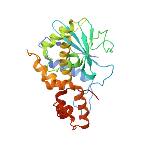
    Maize ribosome-inactivating protein is classified as a class III or an atypical RNA N-glycosidase. It is synthesized as an inactive precursor with a 25-amino acid internal inactivation region, which is removed in the active form. As the first structural example of this class of proteins, crystals of the precursor and the active form were diffracted to 2.4 and 2.5 A, respectively. The two proteins are similar, with main chain root mean square deviation (RMSD) of 0.519. In the precursor, the inactivation region is found on the protein surface and consists of a flexible loop followed by a long alpha-helix. This region diminished both the interaction with ribosome and cytotoxicity, but not cellular uptake. Like bacterial ribosome-inactivating proteins, maize ribosome-inactivating protein does not have a back-up glutamate in the active site, which helps the protein to retain some activity if the catalytic glutamate is mutated. The structure reveals that the active site is too small to accommodate two glutamate residues. Our structure suggests that maize ribosome-inactivating protein may represent an intermediate product in the evolution of ribosome-inactivating proteins.


  • Organizational Affiliation

    Department of Biochemistry, Centre for Protein Science and Crystallography, The Chinese University of Hong Kong, Shatin, N.T., Hong Kong, China.


Macromolecules
Find similar proteins by:  (by identity cutoff)  |  3D Structure
Entity ID: 1
MoleculeChains Sequence LengthOrganismDetailsImage
Ribosome-inactivating protein 3
A, B, C
243Zea maysMutation(s): 0 
Gene Names: b-32CRIP3
EC: 3.2.2.22
UniProt
Find proteins for P25891 (Zea mays)
Explore P25891 
Go to UniProtKB:  P25891
Entity Groups  
Sequence Clusters30% Identity50% Identity70% Identity90% Identity95% Identity100% Identity
UniProt GroupP25891
Sequence Annotations
Expand
  • Reference Sequence
Small Molecules
Binding Affinity Annotations 
IDSourceBinding Affinity
ADE PDBBind:  2PQJ Kd: 6.33 (nM) from 1 assay(s)
Experimental Data & Validation

Experimental Data

  • Method: X-RAY DIFFRACTION
  • Resolution: 2.80 Å
  • R-Value Free: 0.293 
  • R-Value Work: 0.232 
  • R-Value Observed: 0.232 
  • Space Group: P 32
Unit Cell:
Length ( Å )Angle ( ˚ )
a = 115.908α = 90
b = 115.908β = 90
c = 44.898γ = 120
Software Package:
Software NamePurpose
CrystalCleardata collection
MOLREPphasing
CNSrefinement
MOSFLMdata reduction
SCALEPACKdata scaling

Structure Validation

View Full Validation Report


Entry History 

Deposition Data

Revision History  (Full details and data files)

  • Version 1.0: 2008-02-12
    Type: Initial release
  • Version 1.1: 2011-07-13
    Changes: Version format compliance
  • Version 1.2: 2017-08-02
    Changes: Refinement description, Source and taxonomy
  • Version 1.3: 2023-08-30
    Changes: Data collection, Database references, Derived calculations, Refinement description