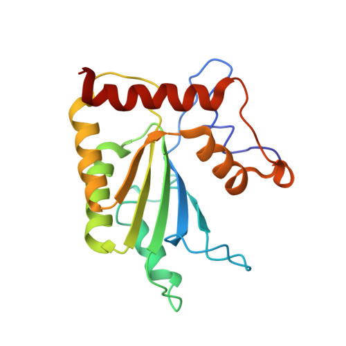
Structure of the Vif-binding domain of the antiviral enzyme APOBEC3G.
Kouno, T., Luengas, E.M., Shigematsu, M., Shandilya, S.M., Zhang, J., Chen, L., Hara, M., Schiffer, C.A., Harris, R.S., Matsuo, H.(2015) Nat Struct Mol Biol 22: 485-491
- PubMed: 25984970
- DOI: https://doi.org/10.1038/nsmb.3033
- Primary Citation of Related Structures:
2MZZ - PubMed Abstract:
The human APOBEC3G (A3G) DNA cytosine deaminase restricts and hypermutates DNA-based parasites including HIV-1. The viral infectivity factor (Vif) prevents restriction by triggering A3G degradation. Although the structure of the A3G catalytic domain is known, the structure of the N-terminal Vif-binding domain has proven more elusive. Here, we used evolution- and structure-guided mutagenesis to solubilize the Vif-binding domain of A3G, thus permitting structural determination by NMR spectroscopy. A smaller zinc-coordinating pocket and altered helical packing distinguish the structure from previous catalytic-domain structures and help to explain the reported inactivity of this domain. This soluble A3G N-terminal domain is bound by Vif; this enabled mutagenesis and biochemical experiments, which identified a unique Vif-interacting surface formed by the α1-β1, β2-α2 and β4-α4 loops. This structure sheds new light on the Vif-A3G interaction and provides critical information for future drug development.
Organizational Affiliation:
1] Biochemistry, Molecular Biology and Biophysics Department, Institute for Molecular Virology, University of Minnesota, Minneapolis, Minnesota, USA. [2] Department of Biochemistry and Molecular Pharmacology, University of Massachusetts Medical School, Worcester, Massachusetts, USA.