2LNK

Solution structure of Ca-bound S100A4 in complex with non-muscle myosin IIA


Experimental Data Snapshot

  • Method: SOLUTION NMR
  • Conformers Calculated: 200 
  • Conformers Submitted: 20 
  • Selection Criteria: structures with the lowest energy 

wwPDB Validation   3D Report Full Report


This is version 1.1 of the entry. See complete history


Literature

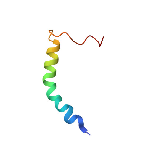
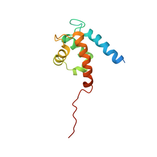
Asymmetric Mode of Ca(2+)-S100A4 Interaction with Nonmuscle Myosin IIA Generates Nanomolar Affinity Required for Filament Remodeling.

Elliott, P.R.Irvine, A.F.Jung, H.S.Tozawa, K.Pastok, M.W.Picone, R.Badyal, S.K.Basran, J.Rudland, P.S.Barraclough, R.Lian, L.Y.Bagshaw, C.R.Kriajevska, M.Barsukov, I.L.

(2012) Structure 20: 654-666

  • DOI: https://doi.org/10.1016/j.str.2012.02.002
  • Primary Citation of Related Structures:  
    2LNK

  • PubMed Abstract: 

    Filament assembly of nonmuscle myosin IIA (NMIIA) is selectively regulated by the small Ca²⁺-binding protein, S100A4, which causes enhanced cell migration and metastasis in certain cancers. Our NMR structure shows that an S100A4 dimer binds to a single myosin heavy chain in an asymmetrical configuration. NMIIA in the complex forms a continuous helix that stretches across the surface of S100A4 and engages the Ca²⁺-dependent binding sites of each subunit in the dimer. Synergy between these sites leads to a very tight association (K(D) ∼1 nM) that is unique in the S100 family. Single-residue mutations that remove this synergy weaken binding and ameliorate the effects of S100A4 on NMIIA filament assembly and cell spreading in A431 human epithelial carcinoma cells. We propose a model for NMIIA filament disassembly by S100A4 in which initial binding to the unstructured NMIIA tail initiates unzipping of the coiled coil and disruption of filament packing.


  • Organizational Affiliation

    Institute of Integrative Biology, BioSciences Building, Crown Street, University of Liverpool, Liverpool L69 7ZB, UK.


Macromolecules
Find similar proteins by:  (by identity cutoff)  |  3D Structure
Entity ID: 1
MoleculeChains Sequence LengthOrganismDetailsImage
Myosin heavy chain, non-muscle IIaA [auth C]39Homo sapiensMutation(s): 0 
UniProt & NIH Common Fund Data Resources
Find proteins for P35579 (Homo sapiens)
Explore P35579 
Go to UniProtKB:  P35579
PHAROS:  P35579
GTEx:  ENSG00000100345 
Entity Groups  
Sequence Clusters30% Identity50% Identity70% Identity90% Identity95% Identity100% Identity
UniProt GroupP35579
Sequence Annotations
Expand
  • Reference Sequence
Find similar proteins by:  (by identity cutoff)  |  3D Structure
Entity ID: 2
MoleculeChains Sequence LengthOrganismDetailsImage
Protein S100-A4B [auth A],
C [auth B]
113Homo sapiensMutation(s): 0 
Gene Names: S100A4CAPLMTS1
UniProt & NIH Common Fund Data Resources
Find proteins for P26447 (Homo sapiens)
Explore P26447 
Go to UniProtKB:  P26447
PHAROS:  P26447
GTEx:  ENSG00000196154 
Entity Groups  
Sequence Clusters30% Identity50% Identity70% Identity90% Identity95% Identity100% Identity
UniProt GroupP26447
Sequence Annotations
Expand
  • Reference Sequence
Experimental Data & Validation

Experimental Data

  • Method: SOLUTION NMR
  • Conformers Calculated: 200 
  • Conformers Submitted: 20 
  • Selection Criteria: structures with the lowest energy 

Structure Validation

View Full Validation Report


Entry History 

Deposition Data

Revision History  (Full details and data files)

  • Version 1.0: 2012-04-25
    Type: Initial release
  • Version 1.1: 2023-06-14
    Changes: Data collection, Database references, Other