2LIP

PSEUDOMONAS LIPASE OPEN CONFORMATION


Experimental Data Snapshot

  • Method: X-RAY DIFFRACTION
  • Resolution: 2.10 Å
  • R-Value Free: 0.220 
  • R-Value Work: 0.163 
  • R-Value Observed: 0.163 

wwPDB Validation   3D Report Full Report


This is version 1.3 of the entry. See complete history


Literature

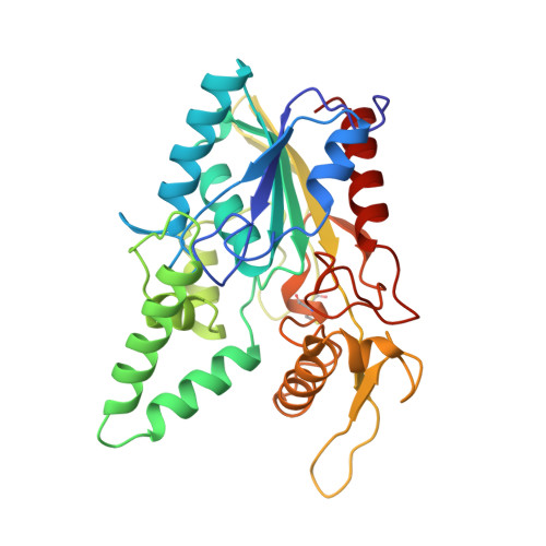
The open conformation of a Pseudomonas lipase.

Schrag, J.D.Li, Y.Cygler, M.Lang, D.Burgdorf, T.Hecht, H.J.Schmid, R.Schomburg, D.Rydel, T.J.Oliver, J.D.Strickland, L.C.Dunaway, C.M.Larson, S.B.Day, J.McPherson, A.

(1997) Structure 5: 187-202

  • DOI: https://doi.org/10.1016/s0969-2126(97)00178-0
  • Primary Citation of Related Structures:  
    2LIP, 3LIP

  • PubMed Abstract: 

    . The interfacial activation of lipases results primarily from conformational changes in the enzymes which expose the active site and provide a hydrophobic surface for interaction with the lipid substrate. Comparison of the crystallization conditions used and the structures observed for a variety of lipases suggests that the enzyme conformation is dependent on solution conditions. Pseudomonas cepacia lipase (PCL) was crystallized in conditions from which the open, active conformation of the enzyme was expected. Its three-dimensional structure was determined independently in three different laboratories and was compared with the previously reported closed conformations of the closely related lipases from Pseudomonas glumae (PGL) and Chromobacterium viscosum (CVL). These structures provide new insights into the function of this commercially important family of lipases.


  • Organizational Affiliation

    Biotechnology Research Institute, NRC of Canada, 6100 Royalmount Ave. Montreal, Quebec H4P 2R2, Canada. Joe.Schrag@nrc.ca


Macromolecules
Find similar proteins by:  (by identity cutoff)  |  3D Structure
Entity ID: 1
MoleculeChains Sequence LengthOrganismDetailsImage
LIPASE320Burkholderia cepaciaMutation(s): 0 
EC: 3.1.1.3
UniProt
Find proteins for P22088 (Burkholderia cepacia)
Explore P22088 
Go to UniProtKB:  P22088
Entity Groups  
Sequence Clusters30% Identity50% Identity70% Identity90% Identity95% Identity100% Identity
UniProt GroupP22088
Sequence Annotations
Expand
  • Reference Sequence
Small Molecules
Ligands 1 Unique
IDChains Name / Formula / InChI Key2D Diagram3D Interactions
CA
Query on CA

Download Ideal Coordinates CCD File 
B [auth A]CALCIUM ION
Ca
BHPQYMZQTOCNFJ-UHFFFAOYSA-N
Experimental Data & Validation

Experimental Data

  • Method: X-RAY DIFFRACTION
  • Resolution: 2.10 Å
  • R-Value Free: 0.220 
  • R-Value Work: 0.163 
  • R-Value Observed: 0.163 
  • Space Group: C 1 2 1
Unit Cell:
Length ( Å )Angle ( ˚ )
a = 91.5α = 90
b = 47.34β = 121.25
c = 85.22γ = 90
Software Package:
Software NamePurpose
RAXISII(MSC)data collection
RAXISII(MSC)data reduction
X-PLORmodel building
X-PLORrefinement
R-AXISdata reduction
R-AXISdata scaling
X-PLORphasing

Structure Validation

View Full Validation Report


Entry History 

Deposition Data

Revision History  (Full details and data files)

  • Version 1.0: 1997-03-12
    Type: Initial release
  • Version 1.1: 2008-03-24
    Changes: Version format compliance
  • Version 1.2: 2011-07-13
    Changes: Version format compliance
  • Version 1.3: 2023-08-09
    Changes: Database references, Derived calculations, Other, Refinement description